L4 Cognition to Contraction IV
 Watch on YouTube →
Overview
Dr. Chris Black explains how calcium released inside a muscle fiber enables actin and myosin interactions in sarcomeres, the structures that generate force. He traces the crossbridge cycle, from calcium shifting troponin and tropomyosin to myosin binding, pulling actin, and using ATP to detach and reset, then connects myosin ATPase differences to muscle fiber types and describes how Bergström needle biopsies let researchers study human muscle.
Key takeaways
- A sarcomere is the basic force-producing unit of muscle: myosin pulls actin toward the center, bringing the Z-discs closer and shortening the unit.
- Calcium activates contraction by binding troponin C, which shifts tropomyosin away from actin's myosin-binding sites; without that shift, the resting state blocks crossbridge formation.
- The crossbridge cycle depends on both ATP hydrolysis and ATP binding: hydrolysis resets the myosin head, while a fresh ATP molecule detaches it from actin.
- Force capacity depends partly on the number of available actin-myosin interactions; the lecture links larger muscle size with more myosin and greater potential force.
- Differences in myosin heavy chains and myosin ATPase activity help distinguish fast-twitch from slow-twitch muscle fibers and relate to contraction speed.
- Bergström needle biopsies provide small, flash-frozen samples of human muscle that researchers can analyze to study muscle structure and fiber types.
Chapters
- The lecture picks up after the cognition-to-contraction signal has traveled through the sarcolemma and T-tubules to trigger calcium release.
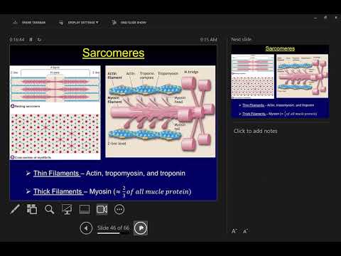
- A muscle fiber contains myofibrils, which are built from sarcomeres, the basic functional units where force is produced and length changes.
- Sarcomeres contain thick myosin filaments and thin actin filaments, the proteins whose interactions drive muscle movement.
- A sarcomere extends from one Z-disc to the next; as it shortens, the Z-discs move closer together.
- Myosin filaments overlap with actin filaments, while a central region contains myosin without actin overlap.
- Myosin heads pull actin toward the sarcomere's center, allowing the filaments to slide past one another and the sarcomere to shorten.
- The lecture sets aside detailed band names such as the A band, focusing instead on filament arrangement and shortening.
- Looking down the sarcomere reveals a hexagonal arrangement, with roughly six actin filaments surrounding each thick myosin filament.
- Myosin heads project at different angles toward surrounding actin, packing many potential interactions into a small space.
- Black describes myosin as about two-thirds of muscle protein and as the molecular motor responsible for force generation.
- A larger muscle generally contains more myosin and has greater potential force capacity, illustrated by comparing the quadriceps with the biceps.
- A myosin molecule has a tail region that contributes to the thick filament and globular heads that interact with actin.
- The lecture distinguishes light meromyosin in the tail from heavy meromyosin in the head region, which contains heavy and light chains.
- Each myosin head has an ATPase enzyme that hydrolyzes ATP and an actin-binding site that forms a crossbridge.
- The head's hinge-like movement transmits force from the molecular scale through sarcomeres and muscle fibers to tendons and bones.
- Tropomyosin lies along the actin filament and blocks myosin-binding sites in the resting state, preventing unwanted contraction.
- Troponin connects to tropomyosin and includes troponin C, which binds calcium released from the sarcoplasmic reticulum.
- Calcium binding changes troponin's shape and shifts tropomyosin away from actin's binding sites, allowing myosin to attach.
- When calcium is removed, the regulatory proteins return toward their resting arrangement and block the binding sites again.
- When myosin binds to an exposed site on actin, the two proteins form a crossbridge.
- A myosin head's hinge movement is the power stroke, which pulls actin and contributes to sarcomere shortening.
- Black demonstrates a biceps curl with an eraser: many crossbridges and power strokes together shorten the muscle and pull on a tendon.
- Crossbridges can form without a visible power stroke during isometric or lengthening actions, topics reserved for the next lecture.
- With calcium exposing actin's binding sites, myosin carrying ADP and inorganic phosphate attaches and forms a crossbridge.
- Phosphate release triggers the power stroke; ADP then leaves, leaving myosin in a lower-energy state.
- ATP binding makes myosin detach from actin, and ATP hydrolysis to ADP plus phosphate resets the head to a higher-energy, ready position.
- Repeated cycles produce sliding filament motion; the lecture emphasizes that ATP binding detaches the crossbridge and hydrolysis recocks the head for another cycle.
- Satellite cells are muscle stem cells located outside the muscle fiber, near its nuclei.
- Black describes the Bergström biopsy needle, developed in Sweden in the 1960s, as a way to collect a small sample of human muscle for cellular study.
- The procedure uses an incision and negative pressure to draw muscle into a needle window, where a cutting cylinder removes a sample about the size of a pencil eraser.
- Researchers can mount and flash-freeze the sample in liquid nitrogen, then prepare sections for later analysis.
- Muscle fiber type is linked to differences in myosin heavy chains and the ATPase activity of myosin heads.
- Researchers can classify fibers as fast-twitch or slow-twitch by examining staining and myosin ATPase function.
- Fast and slow describe the speed of the ATPase system and the fibers' ability to contract and relax; Black connects fast-fiber prevalence with sprinting and jumping ability.
- The lecture closes with a reminder that the cognition-to-contraction assignment is due at 11:59 that night.
Transcript excerpt
0:01 I will have the YouTube channel up today. I added some videos we've done so far on class. I've added the videos yesterday um with the the actual link to that particular playlist did not want to load. I will put that up and get it on in Canvas today. So you guys can go back and watch um go back to watch class if you would like to do so. So here's where we left off on Monday. We were talking about muscles. We had made our cognition to contraction signal. We made our way all the way down and from across the neuromuscular junction down the saroma through a tubule right into the calcium release. So now I'm going to talk about what are
0:48 the smaller sort of cellular microscopic level structures in a muscle that that calcium is going to interact with that's going to get us force production. Okay. So we're going to start off by talking about a myofiberal and then sarcimeir and then within a sarcomir the individual proteins that we're trying to eat this extra protein resistance train to to make more of and So remember inside a muscle p inside the fiber there's a collection of these smaller tubes called myop fibers. So here you have a muscle fiber here. There you can see the cycloplasmic reticulum that's wrapped around in essence if you
1:35 want to say that around that individual myophibbral. So here we're going to pull out one myofiber. Okay, my fibbrals are not that important, but they are made up of something called a sarcimeir. Okay, a sarccomir. And a sarcimeir is the basic functional unit of a muscle. It's at the level of the sarcimeir that we get force production and we get movement. Okay. So, it's the sarcimeir that generates
This is the start of the transcript. Save this video free for the whole transcript, searchable and time-coded.
Related notes
Summary, takeaways, and chapters were generated by AI from the video's transcript and may contain errors. The video belongs to its creator, Exercise Physiology with Dr. Chris Black.