L2 Cognition to Contraction Part II
 Watch on YouTube →
Overview
Dr. Chris Black explains how motor neuron organization and action potentials carry movement commands from the brain through the spinal cord to muscle. He connects spinal motor neuron pools to coordinated recruitment, then traces the electrical signal through resting membrane potential, ion gradients, threshold, depolarization, repolarization, refractory periods, one-way axon conduction, and myelin-assisted speed.
Key takeaways
- Motor neurons supplying the same muscle cluster at related spinal levels, allowing descending commands to recruit the muscle with less timing delay, but making function vulnerable to a spinal injury at those levels.
- A typical neuronal resting membrane potential is about -70 mV, and reaching a threshold near -55 mV triggers an all-or-none action potential.
- Sodium is concentrated outside cells while potassium is concentrated inside; the sodium-potassium pump uses energy to restore these gradients after repeated signaling.
- The absolute refractory period prevents immediate refiring, while the relative refractory period requires a stronger input during hyperpolarization.
- Refractory tissue behind an advancing signal prevents backward propagation, so action potentials normally travel in one direction along an axon.
- Schwann-cell myelin and nodes of Ranvier increase conduction speed; damage to myelin can slow signaling and make movement less coordinated.
Chapters
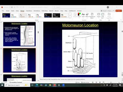
- Motor neurons supplying muscles such as the quadriceps, glutes, hamstrings, and tibialis anterior occupy related spinal levels, illustrated from L4 to S2.
- Clustering motor neurons for one muscle lets descending brain signals reach them at roughly the same time, supporting coordinated recruitment for actions such as sprinting or lifting.
- A spinal cord injury can disable motor neurons below the injury; clustering can therefore mean a muscle group loses function together rather than retaining scattered working neurons.
- In a spinal cord cross-section, the dorsal side faces the back and contains neurons receiving sensory information.
- Motor neurons are located on the ventral side, facing the inside of the body, and their axons leave through spinal nerves toward peripheral muscles.
- The close spinal organization of sensory input and motor output supports integration of information from muscles being recruited.
- An action potential is a brief electrical signal that propagates along specialized nerve and muscle cells.
- A typical resting membrane potential in nervous-system cells is about -70 mV, with the cell interior more negatively charged than the exterior.
- Cells can generate action potentials because they maintain both a membrane voltage difference and unequal ion concentrations across the membrane.
- At rest, sodium concentration is much higher outside a cell, approximately 145 versus 12, while potassium concentration is higher inside.
- Chloride and calcium are also more concentrated outside; closed membrane channels prevent these gradients from immediately dissipating.
- The sodium-potassium pump uses energy to move sodium out and potassium in against their gradients, restoring conditions needed for repeated signaling.
- Maintaining ion gradients can consume a substantial share of resting energy, with estimates reaching roughly one quarter.
- Brain signals descending through the spinal cord reach an alpha motor neuron, where dendrites produce graded changes in membrane voltage.
- As sodium enters, the membrane shifts from about -70 mV toward a threshold near -55 mV; reaching threshold triggers an all-or-none action potential.
- Rapid sodium entry drives depolarization, while slower potassium exit later brings the voltage back down in repolarization.
- Potassium exit can briefly push the membrane below its resting level, causing hyperpolarization before the sodium-potassium pump restores ion concentrations.
- During the absolute refractory period, sodium channels are unavailable, so a second action potential cannot occur regardless of incoming graded signals.
- During the relative refractory period, sodium channels have closed but the membrane is hyperpolarized, so a stronger graded potential is needed to reach threshold again.
- Dr. Black connects hyperpolarization to reliable signaling and explains that voluntary movement typically requires many action potentials, not one.
- A single artificially induced action potential can produce a muscle twitch, while consciously producing exactly one during voluntary movement is impractical.
- An action potential travels from the motor neuron along its axon toward the neuromuscular junction.
- The region immediately behind the advancing signal is in its absolute refractory period, preventing the signal from moving backward and enforcing one-way propagation.
- An electrode can initiate a signal in the middle of an axon, where it can travel in both directions because neither side has just fired.
- Schwann cells produce myelin around peripheral axons; gaps between myelin segments are called nodes of Ranvier.
- High concentrations of sodium and potassium channels at the nodes support rapid signal regeneration, often described as the action potential jumping from node to node.
- Demyelinating conditions such as multiple sclerosis can slow conduction and impair coordination; Dr. Black also recounts a college friend whose immune system attacked axon myelin.
- The lecture ends before explaining neurotransmission at the specialized nerve-muscle synapse, also called the motor end plate or neuromuscular junction.
Transcript excerpt
0:10 I'm really happy. [clears throat] >> Okay. If you don't have a name tag, You don't have a name tag. Come down. >> Got scissors. We've got markers and half and then away we go. Right. >> All right. We will we'll start while you guys are while you guys are doing doing all the Wednesday talking about motor
0:58 neurons talking about the location of motor neurons and where they are contained in the spinal cord which is what we're trying to represent here on um on the screen. Okay. you have collections of motor neurons. So the thousands or tens of thousands of motor neurons that go to your quadriceps or your glutes or your plant flexors or your biceps are all going to be located in a relatively similar place in the spine. So what you can see here is a representation from L4 down to about S2. So we're again put them in the lower lumbar spine and the high sacral spine that's going to be
1:46 here. And these tubes in essence, if you will, are going to represent where all of the motor neurons that are going to be grouped together are going to be. You've got quads just under L4. Got the tibialis anterior. So you've got your what that's a flex. It's a door supply. It's going to do those. You've got your glutes, you got your hamstrings. Okay, so all motor neurons are in kind of roughly the same place with the same muscle. So here's my question for you. Here's my question. You can answer it with answer with somebody sitting next to you. Why do this?
2:34 What's the utility of having all of the motor neurons for same muscle in roughly the same place in the spine? >> Why do you want them spaced out? Oh,
This is the start of the transcript. Save this video free for the whole transcript, searchable and time-coded.
Related notes
Summary, takeaways, and chapters were generated by AI from the video's transcript and may contain errors. The video belongs to its creator, Exercise Physiology with Dr. Chris Black.