L3 Cognition to Contraction Part III
 Watch on YouTube →
Overview
Dr. Chris Black explains how a motor-neuron action potential crosses the neuromuscular junction through acetylcholine, then triggers a muscle action potential and calcium release from the sarcoplasmic reticulum. He connects motor-unit size and muscle-fiber structure to force control, and traces excitation-contraction coupling through T-tubules, dihydropyridine receptors, and ryanodine receptors, identifying this coupling process as a common site of fatigue.
Key takeaways
- At the neuromuscular junction, an axon action potential opens voltage-gated calcium channels; calcium entry triggers acetylcholine release, which activates receptors and starts a muscle action potential.
- The approximately 70-nanometer primary cleft separates nerve from muscle, while secondary membrane folds increase the area available for acetylcholine receptors.
- A motor unit is one motor neuron and all the muscle fibers it innervates; smaller units support precise control in the fingers and eyes, while larger units support high force in muscles such as the quadriceps.
- Muscle force travels through a nested structure of sarcomeres, myofibrils, fibers, fascicles, and whole muscle, with connective-tissue layers carrying force onward to tendons and bones.
- In excitation-contraction coupling, a T-tubule action potential activates dihydropyridine receptors, which open ryanodine receptors and release calcium from the sarcoplasmic reticulum.
- Acetylcholinesterase ends acetylcholine signaling at the neuromuscular junction, while satellite cells provide a muscle-specific stem-cell population involved in repair and growth.
Chapters
- A motor-neuron axon branches near a muscle fiber but does not touch it, leaving a gap that prevents direct electrical transmission.
- Acetylcholine carries the signal across the neuromuscular junction and binds receptors on the muscle membrane.
- The lecture moves from junction structure to the events that occur inside the muscle fiber.
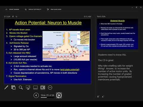
- The axon terminal, or bouton, stores acetylcholine in vesicles and releases it into the primary cleft, an approximately 70-nanometer gap.
- Folds in the muscle membrane form secondary clefts, increasing surface area for acetylcholine receptors.
- Acetylcholinesterase in the basement membrane breaks down acetylcholine, ending the signal; acetylcholine can then be recycled by the nerve.
- A motor unit consists of one motor neuron and every muscle fiber its axon innervates.
- Force increases as the nervous system recruits additional motor units.
- Small motor units in the eyes and fingers support precise force adjustments, while larger units in muscles such as the quadriceps prioritize force.
- An action potential reaching the axon terminal opens voltage-gated calcium channels, and calcium entry triggers acetylcholine-vesicle release.
- Acetylcholine binds muscle-side receptors, opening ion channels and producing a muscle action potential, sometimes called an end-plate potential.
- Acetylcholinesterase terminates receptor activation; repeated nerve impulses sustain the release-and-recycling cycle during movement.
- A skeletal muscle contains many cylindrical muscle cells called muscle fibers, which are unusually large and multinucleated.
- Muscle is mostly water, with roughly 20% protein; actin and myosin are the key force-generating proteins.
- Black uses Russian nesting dolls to introduce the hierarchy of whole muscle, fascicles, muscle fibers, myofibrils, and sarcomeres.
- Actin and myosin are organized in sarcomeres within myofibrils, packing force-generating proteins into muscle fibers.
- Elastic connective tissue links fibers and fascicles so force from individual contractile regions can spread through the muscle.
- The connective-tissue network converges into tendons, transmitting muscle force to bones across joints.
- Epimysium surrounds the whole muscle, perimysium surrounds fascicles, and endomysium surrounds individual muscle fibers.
- These collagen-rich layers provide structural support and help transmit force through the muscle.
- The epimysium contributes to the connective tissue that forms the tendon attaching muscle to bone.
- The sarcolemma is the muscle fiber's membrane; T-tubules carry electrical signals from its surface into the fiber.
- The sarcoplasmic reticulum is an interconnected calcium-storage network positioned alongside the T-tubules.
- Muscle fibers also contain many mitochondria for aerobic ATP production and peripheral nuclei that support protein turnover.
- Satellite cells are muscle-specific stem cells that can be activated to help repair fibers and support muscle growth.
- A muscle action potential travels along the sarcolemma and down T-tubules to the terminal cisternae of the sarcoplasmic reticulum.
- Voltage-sensitive dihydropyridine receptors connect with ryanodine receptors, prompting calcium release into the muscle-cell cytoplasm.
- Black identifies excitation-contraction coupling as a common site of fatigue because impaired signal transmission or calcium release limits force.
- The lecture ends before the next topic, calcium's interaction with sarcomere proteins such as actin and myosin.
Transcript excerpt
0:02 Okay, everybody, if you've got your name tags, please make sure you've got your name tags out so I can fall on all of you if you're already terrified of me probably doing such. We left off on Friday. We had made our signaling pathways all the way down the axons. we were getting very very close to the end very close to where the axons are going to make contact or at the signal contact with the muscle. Okay. So the place where this happens is a very specialized structure called the neuromuscular junction. So we're going to spend today talking about the neuromuscular junction. Then we'll talk about what's going on inside of the muscle fiber
0:50 downstream stuff from the neuromuscular junction. So what you can see in the picture on the left, okay, you can see our motor neuron which is in the spinal cord. You can see our axon that comes off of the motor neuron. You can see the myelin and the nodes of brona. And this is going to project out of the spinal cord all the way out down into a muscle. And then it's going to come into very close contact with an individual muscle pipe. Okay? And it's going to have this highly specialized structure that as you can see here, there's going to be multiple of them coming off of the the axon. But at every given spot on a muscle fiber, it looks like the picture on the right. Okay. So, couple of things to know about
1:38 what's going to happen here. Okay. First, there is no direct contact. The axon comes down, branches, and then sits just right over the top, but it does not quite touch the muscle fiber. So, there's no direct contact. So because of
This is the start of the transcript. Save this video free for the whole transcript, searchable and time-coded.
Related notes
Summary, takeaways, and chapters were generated by AI from the video's transcript and may contain errors. The video belongs to its creator, Exercise Physiology with Dr. Chris Black.