L24 Cardiovascular System and Exercise I
Overview
Chris Black introduces the cardiovascular system as the support network that delivers oxygen, nutrients, and hormones, removes carbon dioxide and waste, and adapts substantially to exercise. The lecture reviews heart anatomy and blood flow, cardiac muscle and electrical conduction, ECG waves, stroke volume and ejection fraction, then defines systolic, diastolic, and mean arterial pressure, including how pressure changes through the circulation.
Key takeaways
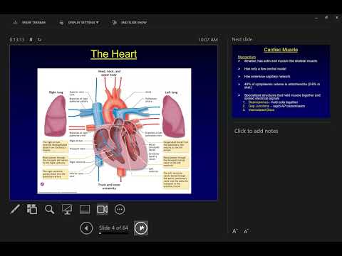
- The heart's blood-flow route is superior vena cava, right atrium, right ventricle, pulmonary arteries, lungs, pulmonary veins, left atrium, left ventricle, and aorta.
- Cardiac muscle contains about 40% mitochondria by cytoplasmic volume, supporting continuous aerobic work, while gap junctions enable electrical signals to spread between cells.
- The SA node initiates each heartbeat, and Purkinje fibers coordinate ventricular contraction from the apex upward to help eject blood.
- Stroke volume is end-diastolic volume minus end-systolic volume; ejection fraction divides stroke volume by end-diastolic volume and can exceed 90% during very intense exercise.
- Mean arterial pressure is estimated at rest as one-third systolic pressure plus two-thirds diastolic pressure, and blood pressure progressively falls from the large arteries toward the right atrium.
Chapters
- The cardiovascular system delivers oxygen, glucose, triglycerides, and hormones to tissues while carrying away carbon dioxide and waste.
- It also contributes to temperature and pH regulation and moves white blood cells, though immune function is not a focus of the lecture.
- Exercise training can adapt the heart, blood vessels, blood volume, and capillary exchange, supporting cardiovascular function as people age.
- The required heart structures are the right and left atria, right and left ventricles, aorta, pulmonary arteries, pulmonary veins, and superior vena cava.
- Blood flows from the superior vena cava to the right atrium and right ventricle, then through the pulmonary arteries to the lungs.
- Oxygenated blood returns through the pulmonary veins to the left atrium and left ventricle, then leaves through the aorta.
- Cardiac muscle, also called myocardium, is striated, has a dense capillary supply, and contains about 40% mitochondria by cytoplasmic volume, compared with roughly 2% to 6% in skeletal muscle.
- Its abundant mitochondria support sustained aerobic metabolism, helping the heart contract continuously without relying primarily on anaerobic metabolism.
- Intercalated discs contain desmosomes that hold adjacent cells together and gap junctions that let ions and electrical signals pass between cells.
- The sinoatrial (SA) node in the upper right atrium spontaneously depolarizes and sets the heart's rhythm; its intrinsic pacing rate is described as about 72 beats per minute.
- The impulse spreads across the atria, reaches the atrioventricular (AV) node, and travels down the AV bundle, also called the bundle of His.
- Purkinje fibers carry activation through the ventricles from the apex upward, coordinating contraction so blood is pushed toward the aorta and pulmonary arteries.
- Heart rate measurements generally reflect ventricular depolarization or contraction, rather than atrial contraction.
- Systole is the contraction phase of a heart chamber; diastole is its relaxation phase, when the chamber can fill with blood.
- Atrial contraction occurs while the ventricles are relaxed, followed by ventricular contraction while the atria fill.
- At roughly 70 to 80 beats per minute, about 40% of the cardiac cycle is systole and 60% is diastole; rising heart rate shortens filling time and brings the phases closer in duration.
- An electrocardiogram records electrical activity across the myocardium using electrodes placed on the body; clinical systems commonly use a 12-lead ECG.
- The P wave represents atrial depolarization, the QRS complex represents ventricular depolarization, and the T wave represents ventricular repolarization.
- Atrial repolarization is obscured by the QRS complex, and the interval between successive R waves shortens as heart rate increases.
- Clinicians assess whether P waves are followed by QRS complexes to evaluate conduction, while ST-segment changes can indicate inadequate blood flow to the heart muscle.
- Stroke volume is the amount of blood ejected from the left ventricle with each beat; a typical resting value is about 80 mL.
- Stroke volume equals end-diastolic volume minus end-systolic volume: the blood present before ventricular contraction minus the blood remaining afterward.
- Ejection fraction is stroke volume divided by end-diastolic volume; a typical resting value is about 66% to 67%, and a low value may indicate impaired ventricular function.
- During exercise, stronger cardiac contraction raises ejection fraction; at very high intensity it can exceed 90%.
- Blood pressure is the force blood exerts on vessel walls and is commonly measured in the brachial artery of the arm.
- The higher reading is systolic pressure during left-ventricular contraction; the lower reading is diastolic pressure during ventricular relaxation.
- Mean arterial pressure is estimated at rest as one-third systolic pressure plus two-thirds diastolic pressure, representing average pressure across a cardiac cycle.
- During exercise, systolic pressure becomes a more prominent driver as heart rate and blood pressure rise.
- Blood moves from higher pressure toward lower pressure, creating the gradient that carries it through the circulatory system.
- Left-ventricular pressure rises sharply during systole and falls very low during diastole; pressure in the large arteries is close to ventricular systolic pressure.
- Pressure progressively declines across arteries, arterioles, capillaries, venules, and veins as blood returns to the right atrium.
Transcript excerpt
0:02 All right. So we finished hormones on I think it was Monday. At least Wednesday. Yeah. We finished hormones on Monday. Now we're going to start talking about the cardiovascular system. And so a lot of what we covered today, I hope we're going to go through pretty quickly, will be review for you guys. Stuff you've seen in anatomy, stuff you've seen in human physiology. Um it should be it should be relatively kind of review stuff. Um I'm going to want you to know even less things structurally here than what you had to know um to get to know in in anatomy class, okay? So then we're going to get into we're going to cover the structures and the basic functions, then we'll get into how all
0:49 of that changes during exercise. That's what you're going to do in lab this week is you're going to you're going to see what happens to heart rate and what happens to blood pressure during exercise, which we're not going to get to cover in in class until probably it'll be Friday or next Monday we'll we'll do all of that, okay? All right. I like to introduce the cardiovascular system by by saying that its primary role is it's support system. And when I say that it's a support system, that doesn't mean that it is secondary in any way, shape or form. In a lot of ways, dysfunction in the cardiovascular system is the leading cause of death in industrialized nations worldwide. And dysfunction in this system will will hit everybody in this classroom in some
1:36 way, shape or form at some point in your life. So we want to understand how things work, what the proper function is so that we can figure out then what exercise does,
This is the start of the transcript. Save this video free for the whole transcript, searchable and time-coded.
In this course
Related notes
Summary, takeaways, and chapters were generated by AI from the video's transcript and may contain errors. The video belongs to its creator, Exercise Physiology with Dr. Chris Black.