091826 Lecture Video
 Watch on YouTube →
Overview
Tim Johann introduces amino acids as protein monomers, explaining how side-chain properties guide protein folding and how the 20 common amino acids are classified and represented. He reviews the structures and one-letter and three-letter codes students need to learn, shows how peptide bonds form, and teaches how to distinguish L and D configurations using spatial orientation and molecular models.
Key takeaways
- In soluble globular proteins, hydrophobic side chains tend to be buried and hydrophilic side chains exposed, reflecting the energetic benefit of reducing ordered water around hydrophobic surfaces.
- The five side-chain groups are nonpolar, polar, aromatic, positively charged, and negatively charged; histidine is mostly unprotonated at pH 7 because its pKa is approximately 6.
- Students are expected to memorize all 20 common amino-acid structures and both their one-letter and three-letter codes for Exam 1.
- Peptide bonds link amino acids through an overall water-elimination reaction, while biological amino-acid addition is coupled to ATP and uses phosphate activation.
- To assign L or D using the taught convention, point the carboxylic acid up and back and the side chain down and back; the amine on the left is L, and on the right is D.
- Protein binding depends on three-dimensional stereochemical fit: an enantiomer can fail to align several functional groups with an enzyme active site even when it has the same atoms.
Chapters
- Quiz material is fair game for the exam, and two years of old exams are available to show question styles.
- Amino acids are the monomers of proteins and can be grouped broadly as hydrophobic or hydrophilic.
- In a soluble globular protein, hydrophobic residues tend to face inward while hydrophilic residues interact with water on the outside.
- This arrangement is driven in part by water entropy, as burying hydrophobic surfaces reduces the need for ordered water structures.
- Most of the 20 amino acids share a backbone with an amine, a carboxylic acid, and a variable side chain.
- The five commonly used side-chain categories are nonpolar, polar, aromatic, positively charged, and negatively charged.
- Proline is structurally distinctive because its side chain connects back to the backbone nitrogen.
- That ring can introduce a kink in a protein chain, affecting how the polymer bends.
- Polar uncharged amino acids include serine, threonine, cysteine, asparagine, and glutamine; several can form hydrogen bonds.
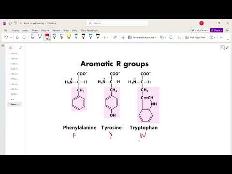
- Phenylalanine, tyrosine, and tryptophan are aromatic, with benzene, phenol, and fused-ring side-chain features, respectively.
- Lysine and arginine are positively charged at pH 7; histidine is mostly unprotonated because its pKa is about 6.
- Aspartate and glutamate are negatively charged; aspartate has one side-chain carbon before its carboxylate, while glutamate has two.
- For Exam 1, students need to memorize the structures of all 20 common amino acids, along with their three-letter and one-letter codes.
- Tim Johann says he will likely provide three-letter codes when testing structure drawing so the question can focus on one concept at a time.
- Many one-letter codes are intuitive, such as glycine G, alanine A, valine V, leucine L, and methionine M.
- Several codes avoid conflicts: asparagine is N, glutamine is Q, tryptophan is W, lysine is K, aspartate is D, and glutamate is E.
- Peptide-bond formation is presented as an overall condensation reaction that eliminates water between amino acids.
- In cells, amino-acid addition is coupled to ATP and uses a phosphate group to create a better leaving group; the board drawing is not the full mechanism.
- Students should be able to draw a peptide bond between specified amino acids, such as aspartate and lysine, and recognize peptide bonds in a larger structure.
- Drawing the backbone in a consistent up-down pattern helps when representing peptide geometry and later hydrogen bonding.
- Proteins interact with molecules across three-dimensional active sites, so a molecule’s stereochemistry determines whether its groups align with the binding surface.
- Tim Johann illustrates the difference using red, green, and black groups: one enantiomer aligns all three, while its mirror image cannot.
- To assign L or D, orient the amino acid with the carboxylic acid pointing up and back and the side chain down and back.
- With that orientation, an amine on the left indicates L and an amine on the right indicates D; cysteine is an exception when converting directly from R/S because sulfur changes priority.
- The class practices rotating drawn structures until the carboxylic acid and side chain point back in the required orientation.
- Adding the hydrogen explicitly as a wedge or dash can make it easier to track the tetrahedral center during rotations.
- The same approach applies to an unusual, non-natural amino acid shown with a cross-linking side chain.
- Students may use molecular model kits during tests and quizzes, and Tim Johann offers to provide models and post a practice worksheet.
- Cyclic peptides can be designed as drugs that block interactions between proteins; Tim Johann recalls a Merck research talk about a peptide aimed at a protein-binding interaction.
- To analyze a residue embedded in a peptide, identify its alpha carbon, amine, carboxylic acid, and side chain before mentally isolating the amino-acid center.
- The example residue is aspartate, recognized by the carboxylic-acid side chain separated from the alpha carbon by one carbon.
- The crowded peptide geometry makes rotations harder to visualize, so a model kit can help track which groups move together.
- The final aspartate example is solved by first positioning the carboxylic acid correctly, then rotating around bonds to place the side chain down and back.
- Tracking the side chain as R, along with the hydrogen and nitrogen, helps avoid swapping groups during each rotation.
- The completed orientation identifies the residue as L-aspartate.
- Tim Johann acknowledges that this is a challenging spatial task and recommends practicing with molecular models; the class ends before covering double pKa topics.
Transcript excerpt
0:01 I'll look unhappy about the quiz. I I'll I'll be grading it later. Uh but uh but all that stuff is fair game on the exam. So we also have two old exams to look at, right? So you have the last two years of exams or maybe there was a skipped here and there, but you have you have two years of old other exams to look at to see kind of how I'm going to ask things. All right. So we're going to start talking about amino acids. Amino acids are the the monomers of proteins. All right. So, we're going to talk about amino acids and proteins, how they go together. Um, there are generally five categories. There are two ways we can break amino acids up into. So, we can break amino acids into two categories or
0:48 five categories. Um the five categories is kind of more traditional what people talk about but in terms of the structure of proteins the the two categories is also very useful. So two categories are are going to be hydrophobic and hydrophilic or polar and non-polar whichever way you want to do this. Okay. And the reason for that hydrophobic and hydrophilic
1:36 division is that if we think about a protein and we think about glob people like to think about like globular proteins, proteins sitting around in solution. Okay, actually it matters for other things. If they're in a membrane as well, you want to think about these sorts of things. But if we just have a free protein floating around in solution, the way that it organizes itself usually is you have the hydrophobic residues on the inside and you have the hydrophilic residues on the outside interacting with water. Okay, so this is the same thing.
This is the start of the transcript. Save this video free for the whole transcript, searchable and time-coded.
Related notes
Summary, takeaways, and chapters were generated by AI from the video's transcript and may contain errors. The video belongs to its creator, Tim Johann.