Cell Injury & Death 2of2 Fall 2026
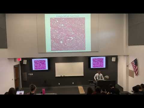 Watch on YouTube →
Overview
Robert Kellar, Ph.D., explains how injury causes distinct patterns of necrosis and traces the cellular mechanisms connecting ischemia, ATP depletion, calcium overload, and reactive oxygen species to tissue damage. Clinical examples include diabetic gangrene, acetaminophen-related liver injury through NAPQI, pigment accumulation, and dystrophic versus metastatic calcification.
Key takeaways
- Coagulative necrosis typically follows ischemia in the heart, kidneys, adrenal glands, and spleen; its wedge-shaped infarcts can heal as nonfunctional fibrotic scars.
- ATP depletion explains several hallmark changes in necrosis: sodium-potassium pump failure causes sodium and water influx, anaerobic glycolysis causes lactic acidosis, and ribosome detachment reduces protein synthesis.
- Excess intracellular calcium activates ATPases, phospholipases, proteases, and endonucleases, damaging energy stores, membranes, the cytoskeleton, and DNA.
- Reactive oxygen species can help macrophages kill bacteria, but uncontrolled oxidation damages cell membranes, proteins, and DNA; antioxidant enzymes limit this injury.
- Acetaminophen is metabolized mostly through sulfate conjugation and glucuronidation, while the cytochrome P450 pathway produces toxic NAPQI that can injure the liver when it accumulates.
- Dystrophic calcification occurs in damaged tissue, whereas metastatic calcification is associated with systemic calcium imbalance such as hypercalcemia.
Chapters
- Necrosis is irreversible and commonly involves cell swelling and a compromised, damaged-looking membrane.
- Apoptosis is controlled cell breakdown involving membrane blebbing, apoptotic bodies, and cell shrinkage.
- Unlike necrosis, apoptosis preserves the plasma membrane and allows cellular components to be recycled.
- Coagulative necrosis commonly affects the heart, kidneys, adrenal glands, and spleen after restricted blood flow causes ischemia.
- Splenic infarcts often have a wedge shape that reflects the branching vascular territory deprived of blood.
- Dead tissue is replaced by fibrotic scar tissue, which lacks the original organ's function; cardiac scarring can compromise rhythm.
- Liquefactive necrosis commonly affects neurons and glial cells, whose breakdown releases hydrolytic enzymes that digest tissue.
- White blood cells, including neutrophils, and tissue macrophages contribute to debris removal and pus formation.
- The abscess-like appearance resembles pus in an infected pimple or splinter, although brain tissue is a key example.
- Caseous necrosis most often appears in lungs affected by tuberculosis, forming yellow-white, cheese-like lesions that combine coagulative and liquefactive features.
- In fat necrosis, lipases break triglycerides into free fatty acids, which bind extracellular calcium, magnesium, and sodium in saponification.
- Saponified fat can form palpable breast nodules, particularly in older, post-menopausal patients, that may resemble cancer on examination.
- Dry gangrene resembles coagulative necrosis: tissue shrinks and turns dark, often in distal extremities with severe hypoxia.
- Unmanaged diabetes can impair small-vessel blood flow to the feet; persistent paresthesia may signal neuropathy and poor perfusion.
- Wet gangrene involves inflammation, neutrophils, pus, and foul odor, while gas gangrene can follow Clostridium infection and gas production.
- Injury sources include hypoxia, trauma, infection, radiation, and chemicals; damage can affect ATP, membranes, the cytoskeleton, or DNA.
- Membrane damage can release cellular enzymes, while cytoskeletal injury disrupts the structural support that maintains cell shape.
- DNA damage can produce misfolded, nonfunctional proteins that prompt programmed cell death.
- Autophagy processes a cell's own components; heterophagy processes material taken up from outside, with both potentially affecting surrounding tissue.
- Hypoxia means low oxygen in tissue; ischemia, or reduced blood supply, is a major cause and can result from vascular disease or trauma.
- Other causes include high altitude, sickle cell disease, cardiovascular or respiratory disease, and respiratory distress such as severe COVID-19.
- Prolonged hypoxia can lower metabolic activity and organelle numbers, causing atrophy; an acute interruption of blood flow can cause necrosis.
- Reduced oxygen limits mitochondrial oxidative phosphorylation, lowering ATP and impairing the sodium-potassium pump.
- Pump failure allows sodium and calcium to accumulate inside the cell; water follows sodium, causing cellular swelling and membrane blebbing.
- The cell temporarily increases anaerobic glycolysis, consuming glycogen and producing lactic acid that lowers intracellular pH.
- ATP loss also detaches ribosomes, reducing protein synthesis, while acidity damages nuclear material and promotes DNA clumping.
- Excess cytosolic calcium can enter from outside the cell or be released from the endoplasmic reticulum and mitochondria.
- Calcium activates ATPases, which consume ATP, and phospholipases, which break down membrane phospholipids.
- Calcium-activated proteases damage cytoskeletal proteins, while endonucleases break down DNA.
- Inflammation, radiation, chemicals, and reperfusion injury can generate superoxide in mitochondria; hydrogen peroxide can then form hydroxyl radicals through the Fenton reaction.
- Macrophages use reactive oxygen species to kill bacteria, but uncontrolled radicals also oxidize lipids, proteins, and DNA.
- Lipid oxidation damages membranes, protein oxidation reduces function, and DNA oxidation can cause mutations.
- Superoxide dismutase, glutathione peroxidase, and catalase help control reactive oxygen species and convert harmful intermediates toward water and oxygen.
- Acetaminophen, the active ingredient in Tylenol, relieves pain and reduces fever but is not an anti-inflammatory drug.
- It is present in many combination products, including DayQuil and NyQuil, so using multiple medicines can unintentionally increase total exposure.
- The liver metabolizes about 90% through sulfate conjugation and glucuronidation; cytochrome P450 processes about 10%, producing the toxic intermediate NAPQI.
- Accumulation of NAPQI damages liver tissue, making careful label reading and attention to patient age and dose important.
- Hemosiderin is an iron-storage pigment that can appear as dark granules in tissue; bruises are a familiar example of iron accumulation.
- Prussian blue staining highlights iron deposits, including punctate hemosiderin in liver tissue.
- Lipofuscin consists of residual material from degraded cellular organelles and accumulates with age, including in cardiac tissue.
- Dystrophic calcification deposits calcium in damaged or dying tissue, as in aortic valves, tuberculosis lesions, and atherosclerotic plaques.
- Metastatic calcification occurs with systemic calcium imbalance, including hypercalcemia associated with hormonal or vitamin D disturbances.
- In the concluding myocardial case, heart tissue and visible early wedge-shaped lesions point to coagulative necrosis.
Transcript excerpt
0:04 What did zero say to eight? Nice belt. What do you call the hair under a cow's nose? A moose. Nicely done. What kind of fruit has the most electricity in it? What kind of fruit has the most electricity in it? A watermelon. All right. The good news is the mic was backwards, so it didn't even get that on the video, so you don't have
0:51 to hear it ever again. Okay. How y'all doing? You ready for a nice long holiday weekend? So, it doesn't really impact us because we're Tuesday, Thursday cadence. So, today you have post quiz two or post quiz one that's due. It's our [clears throat] second week, but it's post quiz one. And um then you have a long weekend. You can work ahead a little bit. Next week we're getting into some, I think, really interesting material, probably some of my favorite material for the for the semester. That's acute and chronic inflammation. Today, [clears throat] we'll kind of set the stage for that.
1:39 So, we're going to we're going to kind of go through the storyline today about the different types of causes of cell and tissue injury. So, we introduced necrosis and we introduced apoptosis. uh we're going to kind of focus now on what happens to the cell, what happens to the tissue when these two types of injurous stimuli show up. So just a quick review, who can share with us some characteristics of necrosis back here? >> Yeah. >> Swelling. What else? Did you have something different? >> It's irreversible. carries in the later stages. >> What about the later stages?
2:28 >> Carries. >> Oh, like dental cavities. Yeah. Yeah. Yeah. For sure. >> Degregation of the membrane or the membrane becomes compromised like it looks motheaten. Remember we showed
This is the start of the transcript. Save this video free for the whole transcript, searchable and time-coded.
Related notes
Summary, takeaways, and chapters were generated by AI from the video's transcript and may contain errors. The video belongs to its creator, Robert Kellar, Ph.D..