092826 Lecture Notes
 Watch on YouTube →
Overview
Protein secondary structures arise from restricted rotations around backbone phi and psi angles, with peptide-bond resonance preventing rotation and Ramachandran plots identifying favored angle combinations. The lecture explains how backbone hydrogen bonds shape alpha helices, how amino-acid placement and charge interactions stabilize them, and how keratin uses coiled coils and disulfide bonds; a 100-residue example shows why exhaustive folding searches across 2^200 possibilities are impractical.
Key takeaways
- Peptide-bond resonance gives the bond partial double-bond character, restricting rotation and leaving phi and psi as the principal adjustable backbone angles.
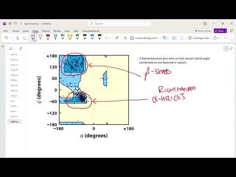
- Ramachandran plots identify favored phi/psi regions, especially those associated with beta sheets and right-handed alpha helices, and can guide structure-modeling penalties.
- A simplified 100-residue protein with two choices for each of 200 backbone angles has about 1.6 × 10^60 conformations, making exhaustive search infeasible even at a hypothetical picosecond per test.
- Alpha helices are stabilized chiefly by hydrogen bonds between backbone amide groups; their 3.6-residue turns also place side chains separated by three to four residues on similar faces.
- Glutamate-rich peptides tend to face electrostatic repulsion above about pH 4, while protonated glutamate groups at lower pH can hydrogen-bond and reduce that destabilization.
- Keratin's right-handed alpha helices assemble into a left-handed coiled coil, and cysteine disulfide bonds help connect larger structures; reducing and reforming those bonds is central to how perms reshape hair.
Chapters
0:00
Protein Secondary Structure and Backbone Rotation Angles
- Secondary structure describes local three-dimensional protein structure, whose elements combine into a protein's global fold.
- Each amino-acid residue is associated with two main rotatable backbone angles, phi and psi.
- These angles, rather than unrestricted rotation at every bond, determine much of the backbone's possible conformations.
2:10
Peptide-Bond Resonance Restricts Backbone Conformation
- Peptide bonds have partial double-bond character because of resonance between the carbonyl and nitrogen.
- That partial double-bond character prevents free rotation around the peptide bond.
- Atoms around each peptide bond lie approximately in a plane, while rotation primarily occurs around the neighboring phi and psi bonds.
5:42
Ramachandran Plots Map Favored Phi and Psi Angles
- A Ramachandran plot places phi and psi angles on its axes and shows which angle combinations occur frequently in proteins.
- Favored regions correspond chiefly to beta-sheet conformations and right-handed alpha helices; left-handed alpha helices are much less common.
- Structure-modeling programs can favor allowed regions with penalties, while permitting unusual angles when experimental restraints support them.
10:42
Why Exhaustive Protein-Folding Searches Fail
- For a simplified 100-amino-acid protein, 200 phi and psi angles with two options apiece yield 2^200, or roughly 1.6 × 10^60, possible conformations.
- Even assuming an unrealistically fast one-picosecond test per conformation, searching the full space would take around 10^40 years.
- Favored backbone geometries and other structural constraints reduce the search space that computational folding methods must consider.
19:33
Alpha Helices and Backbone Hydrogen Bonds
- Alpha helices and beta sheets are identified as the two major protein secondary structures.
- Hydrogen bonds between backbone amide groups are the principal force maintaining secondary-structure shapes.
- The alpha helix discussed is right-handed, with backbone nitrogen and oxygen atoms participating in the hydrogen-bond network.
20:14
Alpha-Helix Turns, Side Chains, Glycine, and Proline
- A typical alpha helix makes one turn every 3.6 amino acids, so residues separated by about three or four positions point toward a similar side of the helix.
- Side chains project outward from the helix, leaving its interior tightly packed rather than forming an open central channel.
- Glycine is often unfavorable because its hydrogen side chain makes it highly flexible; proline is unfavorable because its ring restricts backbone rotation and can bend a helix.
28:40
Helical Wheels Show How Amino-Acid Patterns Stabilize Helices
- A helical-wheel diagram tracks which residues face the same direction, helping identify side chains positioned to interact across roughly three to four residue intervals.
- Nearby opposite charges, such as arginine and glutamate, can attract and contribute to alpha-helix stability.
- Polar and nonpolar residues can also be arranged on different faces, a pattern relevant to helices that associate with other protein regions.
35:47
Glutamate Charge and pH-Dependent Peptide Stability
- A peptide rich in glutamate is expected to become less stable above roughly pH 4, where glutamate side-chain carboxyl groups tend to be negatively charged.
- Repulsion among neighboring negative charges can favor an extended peptide over a compact helical shape.
- Below roughly pH 4, protonated glutamate side chains can hydrogen-bond rather than repel one another, though the transition is gradual and neighboring groups can shift pKa values.
39:43
Applying the Helical Wheel to Build a Stable Alpha Helix
- The exercise uses amino acids including alanine, glycine, leucine, arginine, isoleucine, aspartate, methionine, valine, serine, glutamate, and lysine.
- A workable arrangement places positive and negative residues on compatible faces so opposite charges can interact, while filling other positions with neutral residues.
- Multiple sequences can satisfy the design goal; the key criterion is using the helical wheel to group interacting charges rather than copying one exact sequence.
49:52
Keratin Coiled Coils and Disulfide Bonds in Perms
- Keratin in skin, nails, and horns contains alpha-helical chains that associate as a two-chain coiled coil; the coiled coil is left-handed even though its component alpha helices are right-handed.
- Hydrophobic residues help pack the coiled-coil interior, while hydrophilic residues face outward; larger keratin assemblies are additionally connected by disulfide bonds.
- A disulfide bond links two cysteine side chains; in a perm, chemical reduction loosens these links before hair is reshaped, and reoxidation forms the links again.
Summary, takeaways, and chapters were generated by AI from the video's transcript and may contain errors. The video belongs to its creator, Tim Johann.