100526 Lecture Video
 Watch on YouTube →
Overview
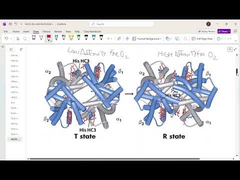
Hemoglobin’s four subunits switch dynamically between the low-oxygen-affinity T state and high-affinity R state, enabling it to load oxygen in the lungs and release it in metabolically active tissues. Oxygen, H+, carbon dioxide, and 2,3-bisphosphoglycerate shift that balance through cooperative structural changes, charge interactions, carbamate formation, and binding in the T-state cavity; increased 2,3-BPG also helps maintain oxygen delivery at high altitude.
Key takeaways
- Hemoglobin’s four subunits cooperate: oxygen binding to one heme pulls its adjacent helix forward and makes oxygen binding at the remaining subunits more favorable.
- The T state has low oxygen affinity and supports tissue delivery; the R state has high affinity and supports oxygen loading in the lungs.
- Low pH favors the T state partly because protonated N-terminal histidine can form a favorable electrostatic interaction with a nearby negatively charged Asp or Glu residue.
- CO2 forms negatively charged carbamates at hemoglobin’s N-termini; their interaction with positively charged lysine residues stabilizes the T state.
- Highly negative 2,3-BPG binds the cavity open in the T state and sterically limits the transition to the R state.
- At high altitude, increasing BPG from roughly 5 mM to 8 mM shifts hemoglobin toward oxygen release, helping offset reduced oxygen loading.
Chapters
0:00
Myoglobin and Hemoglobin: Oxygen Storage Versus Transport
- Myoglobin is a single-subunit oxygen-storage protein, while hemoglobin is an oxygen-transport protein with four interacting subunits.
- Hemoglobin adopts a T state with low oxygen affinity and an R state with high oxygen affinity.
- The T-to-R transition changes the protein’s shape, including the size of the central cavity and the positions of nearby helices.
2:30
Four Factors That Shift Hemoglobin’s Shape
- The four influences discussed are oxygen, H+, carbon dioxide, and bisphosphoglycerate (BPG).
- Oxygen is abundant in the lungs and lower in tissues; H+ and carbon dioxide are higher in metabolically active tissues.
- The bicarbonate buffer system links CO2 removal in the lungs to lower H+; the lecture gives approximate pH values of 7.6 in lungs and 7.2 in tissues.
6:00
Tissues Need Low-Affinity Hemoglobin; Lungs Need High-Affinity Hemoglobin
- Working tissues produce acid and CO2, conditions that favor hemoglobin’s low-affinity T state and oxygen release.
- The lungs expel CO2, lowering H+ and favoring the high-affinity R state for oxygen uptake.
- BPG is present in both lungs and tissues, so it shifts hemoglobin’s overall behavior rather than marking a specific body location.
8:30
Why Hemoglobin Must Switch Affinity States
- A high-affinity-only protein would load oxygen in the lungs but retain it in the tissues; a low-affinity-only protein would load less oxygen.
- Illustrative binding-curve estimates suggest low-affinity hemoglobin could deliver about 30% of its capacity, compared with about 40% when it shifts between states.
- The cooperative state change lets hemoglobin combine strong oxygen loading in the lungs with substantial release in tissues.
13:00
Oxygen Binding Pulls Hemoglobin Toward the R State
- When oxygen binds the iron in one heme, it pulls the helix behind that heme forward.
- Movement in one subunit rearranges the four-subunit protein and pulls corresponding helices in other subunits forward.
- The resulting heme becomes more planar, making oxygen binding at the other subunits more favorable.
17:00
Low pH Favors the T State Through Charge Changes
- Higher H+ concentration at lower pH favors hemoglobin’s T state, reducing oxygen affinity where working tissues need oxygen released.
- Proton binding changes hemoglobin’s charge distribution: some protonatable groups become positive, while some negatively charged groups become neutral.
- The resulting changes disrupt some interactions and create others, shifting the protein toward the T state.
22:00
Protonated Histidine Stabilizes a T-State Salt Bridge
- As tissue pH falls, an N-terminal histidine is more likely to become protonated; the lecture notes that free histidine has a pKa near 6, though a protein environment can shift it.
- In the T state, the positively charged histidine is close to a negatively charged Asp or Glu residue, allowing a favorable electrostatic interaction.
- That interaction is absent when the residues are far apart in the R state, so protonation makes hemoglobin spend more time in the T state rather than locking it into one shape.
28:30
Carbon Dioxide Forms Carbamates That Favor the T State
- In addition to transport as bicarbonate, hemoglobin carries CO2 by reacting it with amino groups at subunit N-termini to form carbamates.
- Carbamate formation gives the N-terminus a negative charge, which can interact with a positively charged lysine on another subunit in the T state.
- Because these groups are farther apart in the R state, CO2 binding stabilizes the T state and promotes oxygen release in tissues.
37:00
2,3-BPG Binds Hemoglobin’s T-State Cavity
- 2,3-bisphosphoglycerate (2,3-BPG) is highly negatively charged and binds in the cavity that is open in the T state.
- Binding in that cavity sterically impedes hemoglobin from closing into the R state until BPG leaves.
- BPG therefore favors the low-affinity T state; the lecture says students should recognize its structure but need not draw it from scratch.
43:00
Higher 2,3-BPG Helps Maintain Oxygen Delivery at Altitude
- The lecture compares about 5 mM BPG at sea level with about 8 mM at high altitude, where lower oxygen availability makes loading more difficult.
- Its illustrative graph estimates roughly 38% oxygen delivery at sea level; at high altitude with sea-level BPG, delivery falls to about 30%.
- Increasing BPG shifts hemoglobin toward the T state: it may reduce oxygen uptake, but increases release to tissues and helps compensate for altitude.
- BPG changes more quickly than red blood cell count, which also rises during longer-term altitude acclimatization.
Summary, takeaways, and chapters were generated by AI from the video's transcript and may contain errors. The video belongs to its creator, Tim Johann.