111126 Lecture Video B
Overview
Tim Johann connects membrane transport to carbohydrate chemistry, then explains how glucose changes between linear and cyclic forms and how its stereochemistry determines structure and stability. He demonstrates Fischer-to-Haworth conversions, glucose ring closure and anomerization, and the formation of alpha-1,4 storage linkages versus beta-1,4 cellulose linkages.
Key takeaways
- Intestinal glucose uptake uses secondary active transport to move glucose uphill into cells, followed by passive transport down its gradient toward the blood.
- Glucose’s cyclic alpha and beta anomers interconvert through a low-abundance linear form; the equilibrium mixture is approximately two-thirds beta, one-third alpha, and 1% open-chain glucose.
- Equatorial substituents reduce steric crowding in a chair conformation, helping make beta-D-glucose more stable and more abundant than alpha-D-glucose.
- Fischer projections encode sugar stereochemistry: D-glucose has the relevant hydroxyl on the right, and its cyclic form places the carbon-5 substituent up.
- The linkage type changes carbohydrate function: alpha-1,4 bonds occur in glucose-storage polymers such as glycogen and starch components, while beta-1,4 bonds build structural cellulose.
Chapters
- Glucose uptake into intestinal cells proceeds against its concentration gradient and requires energy through secondary active transport.
- After glucose accumulates inside the cell, it moves down its gradient toward the blood by passive transport.
- Using energy at only the uptake membrane avoids spending energy on glucose’s downhill movement across the second membrane.
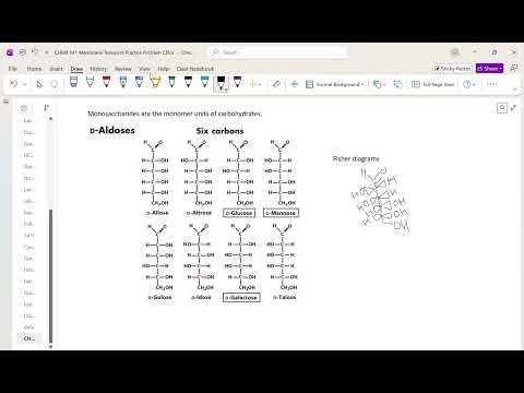
- Monosaccharides are carbohydrate monomers; simple sugars have roughly one water unit per carbon, reflected in their hydroxyl-rich structures.
- Aldoses contain an aldehyde group, while ketoses contain a ketone group.
- Fischer projections encode stereochemistry, and Tim Johann identifies glucose and galactose as the two six-carbon aldoses to memorize.
- Glucose and galactose differ at carbon 4, making them stereoisomers.
- Glucose and galactose can be represented as cyclic Haworth projections as well as linear Fischer projections.
- Cyclization creates the anomeric carbon at carbon 1, which can have either alpha or beta configuration.
- Alpha and beta glucose interconvert in solution by opening to the linear form and closing again.
- The ring-opening and ring-closing process can be accelerated by acid or base; in base, deprotonation creates a stronger nucleophile.
- The nucleophilic hydroxyl attacks the anomeric carbon, forming a six-membered ring that can close in either anomeric configuration.
- At equilibrium, glucose is mostly cyclic: approximately two-thirds beta, one-third alpha, and about 1% in the linear form.
- Acid can promote the reaction by protonating the carbonyl oxygen and making the carbonyl carbon a better electrophile.
- Chair conformations distinguish axial positions, which point up or down, from equatorial positions, which extend outward around the ring.
- Bulky hydroxyl substituents are more stable in equatorial positions because they experience less steric crowding.
- The beta-D-glucose chair places its substituents in especially favorable equatorial positions, helping explain its greater abundance in solution.
- To convert linear glucose to a Haworth projection, number the carbons, map the Fischer left-right pattern to ring up-down positions, and specify alpha or beta at carbon 1.
- For a D sugar, the relevant hydroxyl is on the right in the Fischer projection; in the cyclic representation, the carbon-5 substituent projects up.
- Glucose-derived compounds include glucosamine, a component of chitin, as well as sugars found in glycogen and cellulose.
- Glycogen, amylose, and amylopectin are glucose-based energy-storage polymers; starch contains amylose and amylopectin in proportions that vary by source.
- Glycosidic bonds form when sugars join through dehydration, eliminating water; the linkage is named for the connected carbon positions and anomeric configuration.
- An alpha-1,4 glycosidic bond between glucose units is characteristic of energy-storage polymers such as amylose, amylopectin, and glycogen.
- Cellulose uses beta-1,4 glycosidic bonds, producing a different, structural arrangement of glucose units.
- Diagram conventions vary, but the linkage must clearly show the correct carbon numbers and alpha or beta stereochemistry.
Transcript excerpt
0:05 Okay. All right. What does your answer number three suggest about the relative concentrations of glucose in your intestinal tract compared to outside your cells? Is glucose going up a So, all we care about is a concentration gradient here, right? If we're using energy to bring in glucose, are we going up a gradient or down a gradient here? up, right? Because you'd have you should be going up if uh if you're using energy, right? Does that make sense? Because otherwise you just let you just put a glucon transport there and let that happen. Okay. Okay. So then is energy required to move
0:55 to move glucose across the membrane separating the cell from your blood. Okay. According to the diagram, no, we go up here. We look the glucose just goes through, right? All right. There's no we don't have anything going in. It's just unport. If you just have unapport Okay. Uh well actually that's not going to be a good answer because if you uniport with ATP being being you could have unapport with an ATP being uh being uh hydrayed at the same time. Okay. So the next question is well if we're not using energy and this happens are we going down a concentration gradient or up a concentration gradient?
1:42 >> We have to be going down right? So this is coming down here. All right. And then the final question is probably an explain why or something. Okay. So what explain why energy is only needed to move um glucose across one membrane and not both? Well, if I'm using energy to pump this cell full of glucose, this concentration is higher here than here. always and so it'll come down down down the concentration gradient right so I don't want to use energy here and here because
This is the start of the transcript. Save this video free for the whole transcript, searchable and time-coded.
In this course
Related notes
Summary, takeaways, and chapters were generated by AI from the video's transcript and may contain errors. The video belongs to its creator, Tim Johann.