Placentation in Human Embryology
 Watch on YouTube →
Overview
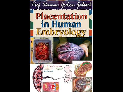
Prof. Akunna Gabriel explains placentation as the progressive formation of the placenta from fetal chorion and maternal decidua, beginning with implantation in week 2 and developing through chorionic villi by week 3. The lecture connects placental structure and function (including exchange, hormone production, and maternal-fetal circulation) to clinical conditions such as placenta previa, placenta accreta spectrum, and preeclampsia.
Key takeaways
- Human placentation begins with implantation in week 2 and develops progressively, with trophoblast differentiation in weeks 2 to 3 and vascularized tertiary chorionic villi forming around week 3.
- The fetal placental component comes from the villous chorion frondosum, while the maternal component comes from the decidua basalis.
- In a hemochorial placenta, maternal blood bathes chorionic villi, but maternal and fetal blood normally remain separate behind the placental barrier.
- The fetal circulation uses two umbilical arteries to carry blood to the placenta and one umbilical vein to return oxygenated blood to the fetus.
- Placental exchange and endocrine functions include nutrient and gas transfer, waste removal, and secretion of hCG, progesterone, estrogen, and human placental lactogen.
- Shallow trophoblast invasion and inadequate remodeling of uterine spiral arteries impair placental perfusion and are associated with preeclampsia.
Chapters
0:00
Implantation and the Timeline of Human Placentation
- Placentation is the progressive formation and functional arrangement of the placenta against the uterine wall, involving fetal chorion and maternal decidua basalis.
- The timeline runs from fertilization and blastocyst formation in week 1 to implantation beginning in week 2, trophoblast differentiation in weeks 2 to 3, and chorionic villus development in weeks 3 to 4.
- The usual implantation site is the posterior-superior uterine wall; the trophoblast contributes to the placenta while the embryoblast forms the embryo proper.
2:20
Trophoblast Layers, Chorionic Villi, and Placental Architecture
- The inner cytotrophoblast supplies cells, while the outer multinucleated syncytiotrophoblast invades the endometrium, interacts with maternal vessels, and produces hCG.
- Chorionic villi mature from primary villi with a trophoblast core, to secondary villi with extraembryonic mesoderm, to tertiary villi containing fetal capillaries around week 3.
- The villi persist in the chorion frondosum to form the fetal placental component; villi regress in the chorion laeve, while the decidua basalis forms the maternal component.
- The early placental barrier includes trophoblast layers, villous connective tissue, and fetal capillary endothelium; it thins as pregnancy advances, affecting transfer of substances and infections.
7:00
Placental Circulation, Functions, and Clinical Abnormalities
- The human placenta is discoid and hemochorial: maternal blood directly bathes chorionic villi, enabling exchange while creating a route for some infections and drugs to cross.
- Maternal blood enters the intervillous space through uterine spiral arteries and drains through uterine veins; two umbilical arteries carry fetal blood to the placenta, and one umbilical vein returns oxygenated blood to the fetus.
- Placental functions include oxygen and carbon dioxide exchange, transfer of glucose, amino acids, and lipids, waste removal, and production of hCG, progesterone, estrogen, and human placental lactogen; IgG transfer supports passive immunity.
- Placenta previa involves implantation in the lower uterus and can cause painless vaginal bleeding; placenta accreta spectrum involves abnormal attachment, while shallow trophoblast invasion and poor spiral-artery remodeling are associated with preeclampsia.
Summary, takeaways, and chapters were generated by AI from the video's transcript and may contain errors. The video belongs to its creator, Prof. AKUNNA Gabriel Lecture Series.