2-4th Week of Development
 Watch on YouTube →
Overview
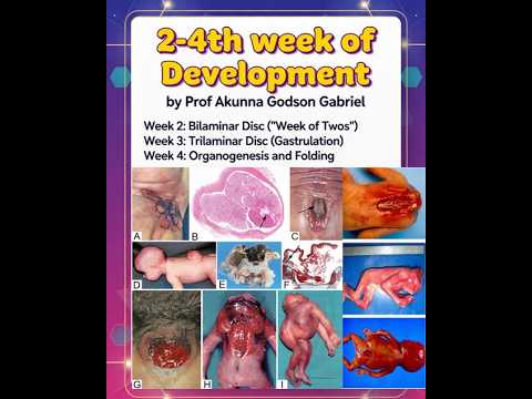
Embryonic development from days 8 to 28 progresses from implantation and the bilaminar disc in week two, through gastrulation and formation of three germ layers in week three, to folding and early organogenesis in week four. The lecture connects these stages to clinical outcomes, including pregnancy-test hCG production, neural tube defects, omphalocele, and early pregnancy loss.
Key takeaways
- Syncytiotrophoblast produces hCG during early pregnancy, providing the hormone measured by pregnancy tests.
- Gastrulation between days 15 and 21 establishes ectoderm, mesoderm, and endoderm, the foundational layers for subsequent tissue and organ development.
- The notochord forms from mesodermal cells and induces the neural plate, which develops into the neural tube.
- Neural tube closure occurs around days 25 to 27; failure can result in anencephaly or spina bifida.
- Week-four lateral folding reshapes the embryo and incorporates the primitive gut; failure of this folding can lead to omphalocele.
Chapters
0:00
Week Two: Implantation, the Bilaminar Disc, and hCG
- During days 8 to 14, implantation completes and the trophoblast differentiates into inner cytotrophoblast and invasive outer syncytiotrophoblast.
- The bilaminar disc consists of epiblast and hypoblast; the amniotic cavity forms within the epiblast, while the yolk sac supports the early embryo.
- Syncytiotrophoblast produces human chorionic gonadotropin (hCG), the hormone detected by pregnancy tests.
- Extraembryonic mesoderm and the chorionic cavity develop around the amniotic cavity and yolk sac.
3:49
Week Three: Gastrulation and the Three Germ Layers
- From days 15 to 21, the primitive streak appears at the caudal end of the epiblast and establishes the craniocaudal axis as gastrulation begins.
- Cell migration forms ectoderm, mesoderm, and endoderm; these layers contribute respectively to structures including the nervous system and skin, muscles and skeleton, and gut lining.
- Mesodermal cells form the notochord, which induces development of the neural plate.
- Placental villi develop in relation to maternal blood, and the connecting stalk contributes to the future umbilical cord; disrupted cell organization can cause defects such as caudal dysgenesis.
5:59
Week Four: Embryonic Folding, Neurulation, and Clinical Risks
- During days 22 to 28, cephalocaudal and lateral folding transform the flat embryonic disc into a cylindrical body and incorporate part of the yolk sac into the primitive gut.
- Neurulation forms the neural tube; the cranial neuropore closes around day 25 and the caudal neuropore around day 27, with failed closure associated with anencephaly or spina bifida.
- Somites arise from paraxial mesoderm and contribute to vertebrae, skeletal muscle, and dermis; the primitive heart tube begins beating around days 22 to 23.
- Lateral-folding failure can cause omphalocele, while errors in early heart development and placental problems can contribute to congenital heart defects and pregnancy loss.
Summary, takeaways, and chapters were generated by AI from the video's transcript and may contain errors. The video belongs to its creator, Prof. AKUNNA Gabriel Lecture Series.