Nuclear impacts on health and medicine 2026 1412 L36
Overview
Dr. Williams explains how radiation dose, exposure duration, and route affect health, then connects radiation protection to medical uses such as technetium-99m imaging and fluorine-18 PET scans. He compares background and medical doses with acute exposures, describes Fukushima Daiichi and workplace monitoring, and challenges the linear no-threshold model, a position presented as his interpretation rather than a settled scientific consensus.
Key takeaways
- Radiation exposure varies widely by source: the lecture estimates background at about 300 millirem per year, while a cardiac catheterization can deliver substantially more than a routine chest X-ray.
- Ionizing radiation can damage tissue indirectly by producing reactive species from water, including hydroxyl radicals that can attack DNA and other organic molecules.
- Radiation protection relies on monitoring and dose minimization: film badges track worker exposure, and ALARA guides efforts to keep doses as low as reasonably achievable.
- Nuclear medicine uses isotope properties for targeted diagnosis: technetium-99m and iodine-123 emit gamma rays for imaging, while fluorine-18 supports PET scans through positron emission.
- PET coincidence detection reduces background interference by accepting paired gamma-ray signals arriving simultaneously at opposing detectors.
- Dr. Williams challenges the linear no-threshold model and presents dose thresholds and biological recovery as central to risk assessment, but his rejection of that model is a contested interpretation rather than established consensus.
Chapters
- Dr. Williams estimates average U.S. background exposure at about 300 millirem per year, from sources including potassium-40 in the body, building materials, soil, and cosmic radiation.
- He notes that radon can substantially increase exposure in areas with uranium-bearing ground.
- A three-hour flight adds a small dose, while cardiac catheterization with continuous X-ray imaging can produce a much larger exposure than a routine chest X-ray.
- Ionizing radiation can transfer enough energy to water molecules to create reactive species, including hydroxyl radicals.
- Hydroxyl radicals can remove hydrogen atoms from organic molecules, potentially damaging DNA, killing cells, or contributing to cancerous changes.
- The lecture contrasts penetrating radiation: paper stops alpha particles, aluminum can stop beta particles, and lead attenuates gamma rays; water can slow neutrons.
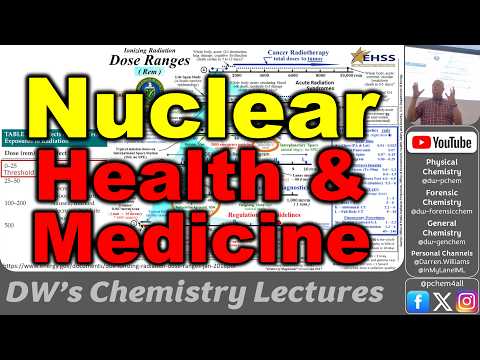
- A Department of Energy dose chart places chest X-rays and dental imaging well below the lecture's cited levels for detectable effects.
- Dr. Williams describes roughly 25 to 50 rem as a range where acute exposure may become detectable through changes such as reduced white blood cell counts.
- LD50/30 means a dose expected to kill 50 percent of an exposed group within 30 days; the lecture places this near 500 rem.
- He contrasts those acute doses with cancer radiotherapy, where high doses are deliberately concentrated on tumors while limiting damage to healthy tissue.
- Film-badge dosimeters record occupational exposure and are collected periodically to measure workers' accumulated dose.
- At Pantex, Dr. Williams says workers were restricted from radioactive areas after reaching 0.1 rem per year, applying the ALARA principle: as low as reasonably achievable.
- He compares this with a nuclear-plant limit of about 0.5 rem per three months and explains that pregnancy-related limits aim to protect developing embryos.
- After the magnitude 9 earthquake in 2011, control rods shut down operating reactors at Fukushima Daiichi, but the subsequent tsunami flooded the plant and disabled backup generators.
- Loss of cooling for spent fuel contributed to overheating, hydrogen production, building explosions, and meltdowns in units 1 through 3; contaminated water also required pumping and treatment.
- Dr. Williams disputes media descriptions of the Fukushima workers as doomed, citing his account that none exceeded 25 rem, while some exceeded administrative limits.
- He argues that public reaction to Fukushima contributed to Germany's decision to phase out nuclear power.
- The lecture compares estimated deaths associated with coal, oil, gas, wind, and nuclear power, emphasizing mining, transport, and installation hazards as well as operational risks.
- Dr. Williams cites an extrapolation of about 10,600 deaths for meeting global energy needs with nuclear power, and contrasts it with much higher figures for coal and oil.
- He offers household falls and automobile deaths as reference points, but the figures are presented as the lecture's comparison rather than a universally established ranking.
- Dr. Williams describes the standard toxicology framework as absorption, distribution, metabolism, and elimination, or ADME, and distinguishes substances that accumulate from those the body clears.
- He argues against the linear no-threshold model, which treats every radiation dose as harmful and additive; this is a contested view, not a scientific consensus established in the lecture.
- He compares radiation damage to a burn, explaining that severe acute exposure can overwhelm the body's ability to repair damaged tissue and may cause acute radiation syndrome.
- The lecture identifies radon inhalation as a particular concern because radioactive material can affect sensitive lung tissue.
- Technetium-99m has a roughly six-hour half-life and emits gamma rays for imaging organs including the heart.
- Iodine-123, with a roughly 13-hour half-life, is taken up by the thyroid and supports thyroid imaging.
- Fluorine-18 is used in PET tracers; after positron emission it becomes oxygen, while the tracer molecule helps reveal biological activity.
- A sodium iodide scintillation crystal converts incoming gamma rays into light flashes, which photomultiplier tubes detect to form an image.
- PET scanners detect pairs of gamma rays produced when a positron meets an electron; coincident signals identify a line through the body and help locate tracer hotspots while rejecting unrelated background events.
- The lecture describes the University of Missouri reactor as a U.S. source of medical isotopes, with neutron-irradiated targets transferred to shielded hot cells for processing.
- Radioactive contamination can be measured with instruments such as Geiger counters, a detection advantage Dr. Williams contrasts with identifying chemical toxins that require sampling.
Transcript excerpt
0:01 Okay, welcome Friday. We made it. We will have class Monday. We'll we'll we'll work on um we'll look at uh nuclear waste and and and all the issues associated with that and compare uh other forms of energy. But then uh for uh after um Thanksgiving break, we'll do um nuclear power. So we'll look at all the different forms of nuclear energy and and new reactor designs and so on. So today we're talking about nuclear medicine and the impacts of radiation on our health. So this is one of the things that people are concerned about. They say, "Oh, radiation is really bad, so we can't do nuclear power or nuclear energy because we're worried about the impacts on our health." When it's probably the opposite. I mean it's one of the safest
0:50 forms of energy and we can uh it actually some of the radionuclides that we get out of the reactors have been transformational for our health and some of the imaging. So let's look at our radiation exposures. You know when I had the little meter up here it was measuring about 13 to 15 counts per minute um in terms of exposure just from the room. And that's because we all have potassium 40 in our bodies. So, we're slightly radioactive and then the concrete and building materials. And so, here's this a little pamphlet from the Department of Energy, and it talks about some of the sources of of our average doses. If we take a three-hour jet plane ride, we get one milligram, which is a really tiny amount of building materials, chest X-rays. The soil has radionuclides in it. Um, and
1:39 then our bodies, we have the potassium 40 in us. Maybe we have some carbon 14 as well from our diet. Um and then uh
This is the start of the transcript. Save this video free for the whole transcript, searchable and time-coded.
In this course
Related notes
Summary, takeaways, and chapters were generated by AI from the video's transcript and may contain errors. The video belongs to its creator, DWs General Chemistry Videos.