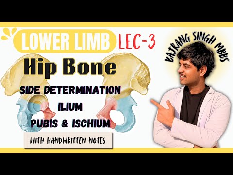
Hip bone anatomy ll osteology, side determination ilium ischium pubis ll Bones of lower limb mbbs
 Watch on YouTube →
Overview
Bajrang Singh explains hip bone osteology, identifying it as the fusion of the ilium, ischium, and pubis around the acetabulum, then demonstrates side determination and key anatomical landmarks. The lesson covers the ilium’s surfaces and borders, the obturator foramen, pubis, and ischium, and recommends the Skeleton 3D app for reviewing bone features.
Key takeaways
- Orient a hip bone by placing the acetabulum laterally, the obturator foramen inferiorly, and the pubis anteriorly.
- The ilium, ischium, and pubis converge at the acetabulum, which articulates with the femur.
- In anatomical position, the ASIS and pubic tubercle align in the same coronal plane.
- Three gluteal lines divide the ilium’s outer gluteal surface into four areas, while its inner surface includes the iliac fossa and sacropelvic surface.
- The obturator nerve, artery, and vein pass through the obturator canal, the opening left where the obturator membrane does not cover the foramen.
- The ischial tuberosity anchors the sacrotuberous ligament, and the ischial spine separates the greater and lesser sciatic notches.
Chapters
- The hip bone, also called the pelvic or coxal bone, forms from the ilium, ischium, and pubis, which meet at the acetabulum for articulation with the femur.
- Determine the side by directing the acetabulum laterally, the obturator foramen inferiorly, and the pubis anteriorly.
- In anatomical position, the anterior superior iliac spine (ASIS) and pubic tubercle lie in the same coronal plane.
- The hip bone articulates with the femur at the acetabulum, with the sacrum at the sacroiliac joint, and with the opposite pubis at the pubic symphysis; sacrotuberous and sacrospinous ligaments help form the sciatic foramina.
- The ilium extends from the iliac crest superiorly to its contribution to the acetabulum inferiorly; the crest ends at the ASIS anteriorly and the posterior superior iliac spine posteriorly.
- Its anterior and posterior borders include the corresponding inferior iliac spines, while the medial border runs from the iliac crest toward the iliopubic eminence.
- The inner surface includes the concave iliac fossa and the sacropelvic surface, which is divided into the iliac tuberosity, an articular surface for the sacrum, and a pelvic surface.
- Three gluteal lines divide the outer gluteal surface into four areas.
- The obturator foramen is bounded anteriorly by the pubis and posteriorly by the ischium; the lesson describes it as typically larger and oval in males and smaller and triangular in females.
- An obturator membrane covers most of the foramen, leaving the obturator canal for passage of the obturator nerve, artery, and vein.
- The pubis has a body, superior ramus, and inferior ramus; the body’s superior border is the pubic crest, whose lateral end forms the pubic tubercle.
- The medial symphyseal surface joins the opposite pubis at the pubic symphysis, while the superior ramus bears the pectineal line and obturator crest.
- The superior pubic ramus extends from the pubic body toward the iliopubic eminence and acetabulum, while the inferior ramus extends from the body to the ischium.
- The ischium contributes to the posterior boundary of the obturator foramen and consists of a body and ramus.
- The ischial tuberosity forms the lower end of the ischium and provides attachment for the sacrotuberous ligament.
- The posterior border contributes to the greater sciatic notch, ischial spine, and lesser sciatic notch.
- Bajrang Singh recommends reviewing the landmarks with notes and the Skeleton 3D app, which can isolate the hip bone for closer inspection.
- The app is used to revisit features including the iliac tuberosity, symphyseal surface, greater and lesser sciatic notches, ischial spine, and obturator foramen.
- The lesson closes by directing learners to a follow-up lesson on the contents of the greater and lesser sciatic foramina.
Transcript excerpt
0:00 Hello everyone, welcome back और अभी हम पढ़ रहे हैं lower limb और lower limb में जो अगला lecture हम पढ़ने वाले हैं वो है hip bone का तो चलो hip bone शुरू करते हैं तो सबसे पहले hip bone जो है hip bone को अपन pelvic bone भी बूलते हैं या फिर इसको हम coaxial bone भी बूलते हैं ठीक है? और hip bone जो है अपने 11 तल्स पे पढ़ाए कि hip bone जो है वो तीन bones के fusion से बनती है ठीक है और वो कौन-कौन सी है पहली ileum फिर ischium फिर pubis ठीक है तो हम देख लेते हैं पहले तो यह पूरी जो bone है अपनी यह पूरी की पूरी hip bone है अपनी और hip bone देखो तुम तो यह तीन bone से बन रह तो Anteriorally Presented Pubis और Posteriorly बट Inferiorly
0:49 Presented On to Postero Inferiorly Presented By तो इस फ़ांथ ऑफ दर फ्री बोंज तरह इस इलियम इसरियम और प्योबिस जिसमें इलियम जो हो Superiorly प्रजेंट है चैनल बोस्त यह पोस्टेर इन्फीरिर्ली प्रजेंट है और प्योभिस जो हो entero-inferiorly present है और यह तीनों की तीनों bones जो है वो fuse हो जाती है और वो बना देती है cavity जिसको हम बोलते है acetabulum और acetabulum के अंदर ही अपनी femur जाके अटाच होती है ठीक है यह देखो यह यहाँ पे आके तीनों की तीनों fuse हो रही है और यहाँ पर एक cavity बनाती है जिसको बोलते थे आशिटा बूलम और अशिटा बूलम देखो यह अशिटा बूलम के अंदर आके यह फीमर अटाच हो जाती है ठीक है तो वही चीज़ है कि all these three joints and forms a cup shaped acetabulum जो articulate करता है फीमर के साथ अगली जो बात है hip bone की वो है कि hip bone जो है वो एक irregularly shaped bone है ठीक है कोई एक इसकी regular shape नहीं है ठीक है तो irregularly shaped है फिर अगर हम कोई भी bone पढ़ते हैं तो सबसे पहले उसके side determination देखते हैं और side determination के अंदर हमें 3 चीज़ देखनी होती है पहला जो इसको differentiate करें कि कौन सा part उपर होगा कौन सा निच्च होगा त
This is the start of the transcript. Save this video free for the whole transcript, searchable and time-coded.
Related notes
Summary, takeaways, and chapters were generated by AI from the video's transcript and may contain errors. The video belongs to its creator, BAJRANG SINGH MBBS.