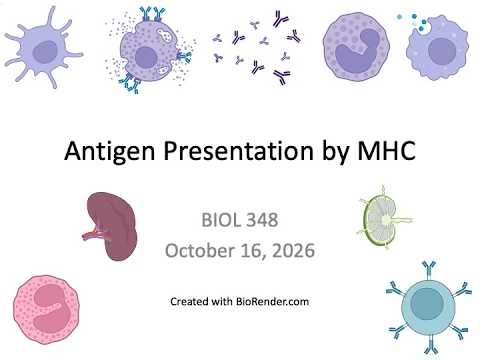
Immunology Fall 2026: Lecture 17 Antigen Presentation by MHC Class I
 Watch on YouTube →
Overview
Brianne Barker explains how MHC class I presents peptides from cytoplasmic proteins to CD8 T cells, organizing the pathway into six stages: acquisition, tagging, proteolysis, delivery, binding, and display. Proteins marked with ubiquitin are cut by the proteasome, peptides cross into the ER through ATP-powered TAP, and tapasin helps position MHC class I for peptide loading; interferon can increase presentation and promote an immunoproteasome that generates more suitable peptides.
Key takeaways
- MHC class I samples cytoplasmic proteins continuously, so healthy cells display self peptides even without infection and infected cells typically display a mixture of self and pathogen peptides.
- Ubiquitin tagging directs cytoplasmic proteins to the proteasome, which produces variable-length fragments rather than specifically generating the 8-to-10-amino-acid peptides favored by MHC class I.
- Type I interferon increases MHC class I presentation and induces immunoproteasome subunits that generate peptides better suited to MHC class I binding.
- TAP uses ATP to move proteasome-generated peptides from the cytoplasm into the ER, while tapasin positions TAP next to MHC class I to improve peptide loading.
- MHC class I display is a cellular quality-control signal: nearly all nucleated cells present it, and loss of display can mark a cell for immune elimination.
Chapters
- Brianne Barker postpones the planned paper discussion and says students should wait for a later announcement about whether it will be eliminated.
- The lecture shifts from MHC structure to the steps that turn intracellular proteins into peptides displayed at the cell surface.
- The ER, Golgi, lysosomes, and transport vesicles are grouped as the secretory pathway, endocytic pathway, or endomembrane system.
- Barker contrasts cytoplasm with compartments across a lipid bilayer, emphasizing that moving material between them requires crossing a membrane.
- Ribosomes begin translation in the cytoplasm, and protein targeting determines whether a protein remains cytosolic or enters the endomembrane system.
- Proteins destined for secretion, the ER, Golgi, vesicles, or the cell membrane are synthesized at the rough ER, using translation-associated energy to move into or across the membrane.
- Transport vesicles bud from one endomembrane compartment and fuse with another while preserving the orientation of lipids and transmembrane proteins.
- During exocytosis, vesicle contents become extracellular, while membrane-protein domains facing the vesicle interior end up facing outside the cell.
- Endocytosis brings extracellular cargo into the endomembrane system, not directly into the cytoplasm.
- MHC class I handles cytoplasmic proteins, while MHC class II handles material associated with the endomembrane system.
- Barker organizes MHC processing into acquisition, tagging for destruction, proteolysis, delivery, binding, and display.
- MHC class I consists of a heavy chain, beta-2 microglobulin, and peptide, and presents to CD8 T cells through the alpha-3 domain.
- MHC class I acquisition is passive: cytoplasmic proteins, including pathogen proteins, are available for processing without a separate antigen-uptake step.
- Cellular proteins have limited half-lives and are continually replaced, so normal self-proteins as well as pathogen proteins enter the degradation pathway.
- A chain of four ubiquitins with the appropriate linkage marks a protein for destruction, functioning as a molecular signal for the proteasome.
- The proteasome recognizes ubiquitinated proteins, removes and recycles ubiquitin, unfolds the target, and cuts it into peptides inside a catalytic barrel.
- Proteasomes do not specifically produce only the 8-, 9-, or 10-amino-acid peptides preferred by MHC class I; they generate fragments of many lengths.
- During infection, type I interferon induces immunoproteasome subunits that improve the supply of peptides suitable for MHC class I presentation.
- MHC class I is synthesized in the ER because it is a transmembrane protein destined for the cell surface, with its peptide-binding cleft oriented into the ER lumen.
- Proteasomal peptides are in the cytoplasm while the MHC class I binding cleft is in the ER, creating a delivery problem across the membrane.
- TAP, the transporter for antigen processing, uses ATP to pump short peptides from the cytoplasm into the ER.
- Tapasin links TAP to the open MHC class I molecule, positioning the binding cleft near incoming peptides.
- Other ER chaperones help stabilize MHC class I and can assist with peptide trimming when fragments are too long.
- The peptides transported by TAP reflect whatever is being degraded in the cytoplasm, so they can include both self and pathogen-derived peptides.
- Only peptides with compatible anchor residues bind a given MHC class I molecule; the cleft does not accept every peptide.
- Once a suitable peptide stabilizes the MHC class I complex, it proceeds through the secretory pathway to the cell surface.
- Barker describes surface display as constitutive after successful folding and peptide loading.
- Healthy cells continuously present self peptides generated through ordinary protein turnover; presentation does not begin only after infection.
- Infected cells can display a mixture of self-derived and pathogen-derived peptides, and interferon can increase MHC class I presentation.
- Nearly all nucleated cells present MHC class I, whereas red blood cells are an exception; loss of class I display signals a problem and can lead to cell killing.
- The animation traces viral proteins and normal cellular proteins through proteasomal cleavage, TAP transport, ER loading, and cell-surface display.
- It shows calnexin and ERP57 assisting heavy-chain folding, beta-2 microglobulin supporting groove formation, and calreticulin joining the peptide-loading complex.
- Tapasin stabilizes the loading complex, connects MHC class I to TAP, and favors high-affinity peptide loading before the complex travels to the surface.
- The animation concludes that CD8 T cells can recognize viral peptide-MHC class I complexes and kill infected cells using perforin and granzymes.
- Barker schedules the MHC class II pathway for Monday and ends with Shiv Pillai's immunology rap, which repeatedly highlights TAP.
- The rap also previews course topics including lymphocyte trafficking, dendritic cells, cytokines, antibodies, and cytotoxic T cells.
Transcript excerpt
0:00 All right. Um, I'm going to go ahead and get started. Um, first of all, just so you know, I'm in grading jail this weekend. Um, so that's my plans. Um, so be ready for after. Um some of you um, reached out in the original schedule uh, for the course. I had originally had a paper discussion for today. Um, if you look at the updated schedule that I posted on September 28th, um, I pushed that back. Um, looks like maybe I didn't hit the get the deadline on the Moodle submission type. It's okay. This is like the first time I've been using Moodle as much in that way and so I sometimes forget that I have to like go back in
0:47 and change those deadlines. Um it's all right, that's it's a solvable problem. Um I actually have been sort of trying to decide what I want to do about that paper, um, cuz I'm a little bit behind and so I might actually eliminate that paper. Um, I'll probably make that decision after I see how far I get today. Um, so I will let you know about um, what might be going on with that. Um, so um, until sort of future um, announcement, don't worry about that paper. I'll let you know. Um so today we're going to continue talking about um, antigen presentation um, by MHC. Um we talked a lot about the actual MHC
1:34 molecule um, itself um, that we see here on the surface of the cell presenting peptides to um, a T cell receptor in order to activate T cells. Um, but really what we need to focus on um, now is sort of this earlier part of the process, how we go from this protein in the cell
This is the start of the transcript. Save this video free for the whole transcript, searchable and time-coded.
Related notes
Summary, takeaways, and chapters were generated by AI from the video's transcript and may contain errors. The video belongs to its creator, Brianne Barker.