Inflammation 2of2 Fall 2026
 Watch on YouTube →
Overview
Robert Kellar explains how inflammatory mediators coordinate acute inflammation, tracing arachidonic acid into prostaglandins, prostacyclins, thromboxanes, and leukotrienes, and connecting complement and coagulation to immune-cell recruitment. He contrasts acute inflammation, which usually resolves within two weeks, with chronic inflammation marked by persistent macrophages, foreign-body giant cells, and fibrosis, then presents research on UV-photolinked, fibronectin-coated polypropylene mesh that reduced giant-cell counts by nearly half after two weeks in a rodent implant model.
Key takeaways
- Phospholipase releases arachidonic acid from cell membranes; cyclooxygenase produces prostaglandins, prostacyclins, and thromboxanes, while 5-lipoxygenase produces leukotrienes.
- Aspirin and COX inhibitors block the cyclooxygenase branch but leave leukotriene signaling intact; corticosteroids act upstream by suppressing phospholipase activity.
- Complement activation converges on C3 and supports inflammation through leukocyte recruitment, enhanced phagocytosis, and membrane attack complex-mediated lysis.
- Acute inflammation usually resolves within two weeks, but wound remodeling can continue for about a year; chronic inflammation can instead sustain macrophages, fibrosis, and tissue dysfunction.
- Multinucleated foreign-body giant cells and a collagen-rich fibrous capsule are key histological signs of chronic inflammation around implanted materials.
- In a rodent model, UV-photolinked human fibronectin coating on polypropylene mesh reduced foreign-body giant cells by nearly half and significantly reduced fibrous-capsule area after two weeks.
Chapters
0:00
Inflammation Review: Acute and Chronic Types, Exudate, and Classic Signs
- The class reviews acute and chronic inflammation as innate, nonspecific defenses that are necessary for survival.
- Vasodilation and vessel leakage produce fluid-rich transudate or protein-rich exudate.
- Pain, redness, swelling, and heat are the classic signs of inflammation.
4:23
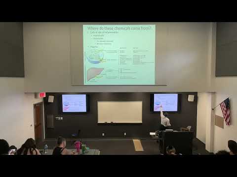
Where Inflammatory Mediators Come From: Injured Tissue, Leukocytes, and Plasma
- Damaged tissue releases mediators such as histamine, which promotes vasodilation and helps initiate leukocyte recruitment.
- Leukocytes leave vessels through margination, rolling, adhesion, and transmigration, also called diapedesis.
- Mediators may be preformed and released from cellular granules or synthesized de novo at the site of inflammation.
- Plasma-borne mediators include liver-produced complement proteins and factors in the coagulation and kinin systems.
12:16
Arachidonic Acid and the Four Eicosanoid Families
- Phospholipase releases arachidonic acid from cell-membrane phospholipids, providing the starting material for 20-carbon eicosanoids.
- Cyclooxygenase enzymes produce prostaglandins, prostacyclins, and thromboxanes; these affect vessel tone, permeability, and platelet aggregation.
- The 5-lipoxygenase pathway produces leukotrienes, which can cause bronchospasm, vasoconstriction, and increased vascular permeability.
18:14
How Aspirin, COX Inhibitors, and Corticosteroids Suppress Inflammation
- Aspirin and COX-1 or COX-2 inhibitors block cyclooxygenase products but do not block leukotriene production.
- Corticosteroids act farther upstream by inhibiting phospholipase activity and reducing arachidonic-acid production.
- Local steroids can target tissues such as the lungs or an arthritic joint; systemic steroid courses are tapered because prolonged suppression can impair immune defense.
22:33
Complement Pathways: C3, Phagocytosis, and Membrane Attack
- The alternative, classical, and lectin complement pathways converge on C3 activation.
- Complement promotes leukocyte recruitment and increases the efficiency of phagocytosis.
- The membrane attack complex assembles in microbial membranes and can cause cell lysis.
25:35
Factor XII Connects the Kinin and Coagulation Systems
- Activated factor XII, also called Hageman factor, initiates both the kinin system and the clotting cascade.
- Bradykinin promotes vasodilation, while the clotting cascade converts prothrombin to thrombin and fibrinogen to fibrin to form a clot.
- Controlled stasis helps move leukocytes toward vessel margins; interference with coagulation can reduce monocyte recruitment.
- Kellar's summary groups vasoactive amines, eicosanoids, complement, and kinin/coagulation mediators by their inflammatory functions.
30:54
Acute Wound Healing Versus Persistent Chronic Inflammation
- Acute inflammation usually resolves in under two weeks, though aging and comorbidities can prolong the response.
- Healing phases overlap: inflammation is followed by cell proliferation and matrix deposition, then remodeling that can continue for about a year.
- Kellar describes retearing his triceps three times before an MRI showed an approximately 97% tear, followed by surgery on January 5.
34:18
Chronic Inflammation in Tissue: Macrophages, Fibrosis, and Lung Changes
- Unlike the transient neutrophil and macrophage response in acute inflammation, chronic inflammation can maintain elevated macrophages and promote new blood vessels.
- Macrophages can fuse into multinucleated foreign-body giant cells, a hallmark of chronic inflammation.
- Persistent inflammation can cause fibrosis and metaplasia; Kellar contrasts these changes in chronic lung tissue with neutrophil-rich acute pneumonia.
- Chronic damage often heals by repair and scar formation rather than full regeneration, leaving functions such as hair growth or normal skin elasticity impaired.
41:12
Chronic Inflammation Histology: Giant Cells and Fibrous Capsules
- Histology shows multinucleated foreign-body giant cells and macrophages around a chronic inflammatory core.
- A fibrous capsule forms as collagen accumulates around the affected area; giant cells and capsule formation distinguish chronic from acute inflammation.
- Macrophage-derived TNF and interleukin-1, together with lymphocyte-derived interferon-gamma, can sustain a self-perpetuating inflammatory cycle.
45:03
Fibronectin-Coated Polypropylene Mesh and Implant Inflammation Research
- Polypropylene is widely used in sutures and meshes for procedures such as hernia repair, but a rodent implant study showed a strong foreign-body giant-cell response after two weeks.
- Kellar describes applying human fibronectin to polypropylene mesh using UV photolinking, which covalently bonds the coating to the surface.
- In the two-week comparison, coated mesh had nearly half as many foreign-body giant cells per fiber as the uncoated control.
- Digital slide analysis also found a statistically significant reduction in fibrous-capsule area, supporting the coating as a way to make the implant more biocompatible.
Summary, takeaways, and chapters were generated by AI from the video's transcript and may contain errors. The video belongs to its creator, Robert Kellar, Ph.D..