Adaptive Immunity 2of2 Fall 2026
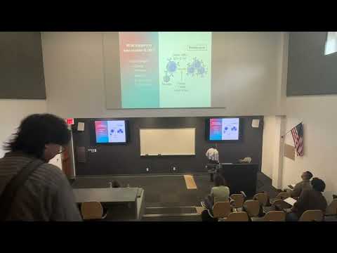 Watch on YouTube →
Overview
Robert Kellar reviews adaptive immunity for an upcoming 50-question exam, covering B-cell antibody functions and memory, T-cell tolerance, antigen presentation, and cytotoxic responses. He connects these mechanisms to vaccination and uses practice questions—including C3b-mediated opsonization and Factor XII activation—to reinforce key concepts.
Key takeaways
- Antibodies can act directly by neutralizing, agglutinating, or precipitating antigens, and indirectly by enabling C3b-mediated opsonization or complement-driven inflammation.
- A primary antibody response takes several days and begins mainly with IgM; secondary exposure produces a much faster response with substantially higher IgG levels.
- T-cell tolerance operates centrally in the thymus and peripherally through apoptosis, anergy caused by missing co-stimulation, and regulatory T-cell suppression.
- MHC class II presentation by antigen-presenting cells primarily activates CD4 helper T cells, whereas MHC class I presentation by infected body cells directs CD8 cytotoxic T cells to their targets.
- C3b promotes phagocytosis by opsonizing microbes, while Factor XII (Hageman factor) connects contact activation to both coagulation and the kinin system.
- Memory T cells include lymph-node-associated central memory cells and tissue-homing effector memory cells, with markers such as CD62L and CCR7 helping distinguish their trafficking patterns.
Chapters
0:00
Exam Format and Adaptive Immunity Foundations
- The online exam has 50 multiple-choice questions, lasts one hour, and is available from 7 a.m. to 11:59 p.m. Tuesday.
- Image questions will use lecture material, with roughly six images expected; Monday office hours are available in person at Wetaw 211 and on Zoom.
- Adaptive immunity is antigen-specific and develops memory; B cells mature in bone marrow, while T cells mature in the thymus.
- Dendritic cells, macrophages, and B cells are antigen-presenting cells that display antigens through MHC molecules.
5:58
B-Cell Central Tolerance and Antibody Effector Functions
- The bone marrow screens developing B cells for self-reactivity; autoreactive cells can be deleted or undergo receptor editing before entering circulation.
- Naive B cells express IgM and IgD, and helper T-cell signals support their proliferation and antibody production.
- Direct antibody effects include neutralization, agglutination (clumping), and precipitation of antigens out of solution.
- Indirect effects include C3b-assisted opsonization and phagocytosis, plus complement-driven inflammation that recruits neutrophils.
13:50
B-Cell Memory, Antibody Responses, and Vaccination
- A primary antibody response peaks after roughly three to five days, with IgM appearing first and IgG following.
- On secondary exposure, IgG levels can be about four times higher than in the initial response, while IgM levels are described as similar.
- Edward Jenner’s 1796 work using cowpox to protect against smallpox helped establish vaccination; the term derives from the Latin word for cow, vacca.
- Influenza and COVID-19 vaccination must account for viral mutation, which can change antigens and reduce how well existing immune memory matches new strains.
19:30
T-Cell Development and Peripheral Tolerance
- Bone-marrow-derived T-cell precursors mature in the thymus, where thymopoietin supports T-lymphocyte maturation and clonal diversity develops.
- Central tolerance removes many self-reactive T-cell clones through apoptosis; Kellar notes that roughly 95% of developing T cells are eliminated.
- Peripheral T-cell tolerance includes apoptosis, anergy when co-stimulation is absent, and suppression by regulatory T cells.
- Unlike the lecture’s simplified contrast with B cells, B cells can also be regulated outside the bone marrow; their tolerance mechanisms are not limited exclusively to central screening.
23:21
MHC Class II and Class I Routes to Helper and Cytotoxic T Cells
- Dendritic cells carry antigens from peripheral tissues into lymph nodes and commonly present them on MHC class II to CD4-positive helper T cells.
- Activated CD4 T cells can promote macrophage activity and cytokine-driven inflammation at infection sites.
- Infected body cells present intracellular antigens on MHC class I to CD8-positive T cells.
- CD8 T-cell activation involves IL-2 signaling before cytotoxic cells leave the lymph node and target infected cells.
26:23
CD8 Cytotoxic Killing and Natural Killer Cells
- Cytotoxic CD8 T cells recognize antigen presented on MHC class I and trigger apoptosis in infected target cells.
- Perforin and granzymes help initiate target-cell apoptosis; Fas ligand binding to Fas is another apoptotic route.
- Natural killer cells are a distinct lymphocyte population, not a subtype of CD8 T cells; the lecture estimates they make up 5–20% of circulating lymphocytes.
- NK cells can use perforin and granzymes to kill abnormal cells without relying on the same antigen-specific recognition used by CD8 T cells.
30:51
Central and Tissue-Homing Memory T-Cell Populations
- Prior antigen exposure produces antigen-experienced T cells that mount a faster, stronger response on re-exposure.
- Central memory T cells tend to remain associated with lymph nodes, while effector memory cells circulate through peripheral tissues.
- Markers discussed include CD62L, CCR7, and IL-7 receptor; lower CD62L and CCR7 are associated with tissue-homing effector memory cells.
- Memory populations divide immune surveillance between lymph-node monitoring and direct patrol of peripheral tissues.
33:52
Practice Questions: Opsonization, Necrosis, and Factor XII
- The pairing review links CD4-positive T cells with MHC class II and CD8-positive T cells with MHC class I; fibroblasts are not standard professional antigen-presenting cells.
- Mast-cell prostaglandins and leukotrienes support inflammation by promoting vasodilation and increased vascular permeability.
- C3b is the substance that enhances leukocyte engulfment of bacteria by acting as an opsonin.
- Factor XII, also called Hageman factor, initiates both the coagulation and kinin systems after contact with an injured vascular basement membrane.
Summary, takeaways, and chapters were generated by AI from the video's transcript and may contain errors. The video belongs to its creator, Robert Kellar, Ph.D..