101926 Lecture Video
 Watch on YouTube →
Overview
Tim Johann connects vitamin C deficiency to weakened collagen: hydroxylation of proline helps stabilize collagen, and ascorbate keeps the prolyl hydroxylase iron cofactor active. The lecture then reviews serine protease catalysis, amino-acid D/L chirality, multi-pKa protonation and Henderson-Hasselbalch calculations, ending with exam guidance on turns and enzyme kinetics.
Key takeaways
- Vitamin C supports collagen formation by keeping prolyl 4-hydroxylase iron reduced; without ascorbate, enzyme inactivation limits hydroxyproline formation and weakens collagen stability.
- Hydroxyproline increases the stated collagen melting temperature from about 41°C to 69°C, well above the 37°C body temperature.
- Serine proteases use serine for covalent catalysis and histidine for acid-base catalysis; water-mediated deacylation releases the product and restores the active site.
- Aspartate stabilizes protonated histidine in the serine protease catalytic triad, shifting histidine’s pKa from roughly 6 to 12.
- At pH 9.25, the illustrative lysine backbone amine with pKa 10 is about 85% protonated and 15% deprotonated, as predicted by Henderson-Hasselbalch.
- Hydrogen bonds stabilize turns, glycine contributes flexibility, and proline promotes the bend.
Chapters
0:00
Collagen’s Gly-X-Y Sequence and Hydroxyproline Stability
- Collagen chains form individual left-handed helices that assemble into a right-handed triple helix.
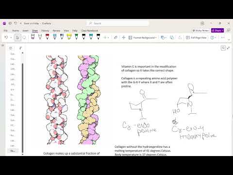
- The repeating sequence is Gly-X-Y, with proline often appearing at X and Y; hydroxylation produces 4-hydroxyproline and changes the ring geometry.
- The lecture compares collagen melting temperatures of about 41°C without hydroxyproline and 69°C with it, illustrating how hydroxylation stabilizes connective tissue.
7:45
Vitamin C, Prolyl Hydroxylase, and Scurvy
- Prolyl 4-hydroxylase uses alpha-ketoglutarate and an iron cofactor to hydroxylate proline residues in collagen.
- Ascorbate, the active form of vitamin C, helps keep the enzyme’s iron in the reduced Fe2+ state; without it, iron becomes Fe3+ and the enzyme is inactivated.
- Insufficient hydroxyproline lowers collagen stability near the 37°C body temperature, helping explain scurvy’s connective-tissue weakness and bleeding symptoms.
14:06
Serine Protease Catalysis: Acylation of the Peptide Substrate
- A serine protease uses histidine as an acid-base catalyst and serine as a nucleophile to cleave a peptide bond.
- Histidine removes a proton from serine, enabling serine to attack the substrate carbonyl and form a covalent enzyme-substrate intermediate.
- The carbonyl reforms as the peptide bond breaks; histidine donates a proton to the departing nitrogen-containing product.
19:00
Serine Protease Deacylation and Histidine’s Shifted pKa
- Water enters the active site, histidine removes a proton, and the resulting hydroxyl attacks the enzyme-bound carbonyl to release the second peptide fragment.
- The serine, histidine, and aspartate catalytic triad supports covalent and acid-base catalysis, then regenerates the enzyme.
- Aspartate’s negative charge stabilizes protonated histidine, raising its pKa from about 6 to roughly 12 and making it a stronger base in the active site.
22:50
Mechanism Drawing Rules and Approximate pKa Values
- For serine protease mechanisms, focus on the proton transfers and covalent bond changes; catalytic-site hydrogen bonds can be omitted from the arrow-pushing sequence.
- Approximate values to remember are pKa 2 to 4 for carboxylic acids and 9 to 10 for amines.
- Tim Johann recommends reviewing practice tests and worksheets because serine protease mechanisms appear in previous exams.
24:30
Assigning D and L Amino-Acid Configuration
- Orient the amino acid with the carboxyl group at the top and the side chain at the bottom; an amine on the left indicates L, while an amine on the right indicates D.
- A molecular model or careful three-dimensional rotation can help convert wedge-and-dash drawings into the required orientation.
- The lecture connects D/L labels with R/S assignments while noting cysteine as an exception, and recommends practicing with the posted worksheet.
30:13
Mapping Lysine’s Three pKa Values to Its Functional Groups
- The worked lysine example assigns illustrative pKa values of 4 to the backbone carboxyl group, 10 to the backbone amine, and 12 to the side-chain amine.
- At pH 9.25, the carboxyl group is deprotonated and the side-chain amine remains protonated; the backbone amine is the group with two significant forms.
- Using the lecture’s 1% rule, groups more than about two pH units from their pKa are treated as having only one relevant protonation state.
38:02
Henderson-Hasselbalch Calculation for Lysine at pH 9.25
- For the backbone amine, the example applies pH = pKa + log(A−/HA), using pH 9.25 and pKa 10.00.
- The calculated ratio A−/HA is about 0.1778; combining the forms gives approximately 85% HA and 15% A−.
- A consistency check confirms that pH below pKa should favor the protonated acid form, matching the larger HA percentage.
43:29
Turn Stability and Final Exam Topics
- Protein turns are stabilized primarily by hydrogen bonds; glycine adds conformational flexibility, while proline helps induce a turn.
- Students are expected to understand that beta turns exist and what stabilizes them, but not necessarily to draw a beta-turn structure.
- Tim Johann confirms that reaction-coordinate diagrams and Michaelis-Menten material may appear on the test.
Summary, takeaways, and chapters were generated by AI from the video's transcript and may contain errors. The video belongs to its creator, Tim Johann.