101429 Lecture Video
 Watch on YouTube →
Overview
Tim Johann explains how to calculate enzyme activity from an NADH absorbance slope, using the Beer-Lambert law, dilution correction, reaction volume, and enzyme volume to derive a total activity of about 65 units. He then connects protein shape changes to induced fit, allosteric regulation, and local pKa shifts before explaining how misfolded prion proteins can propagate their shape and cause diseases such as Creutzfeldt-Jakob disease.
Key takeaways
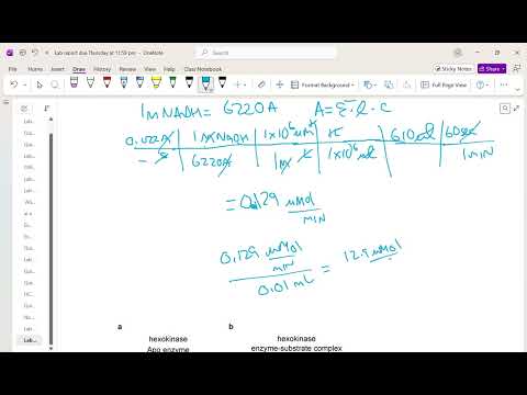
- For the example assay, a 0.022 absorbance-unit-per-second NADH slope, a 6,220 M⁻¹ cm⁻¹ extinction coefficient, a 610 µL reaction, and a 10 µL enzyme aliquot yield about 0.129 µmol/min in the tube and about 65 total units across 5 mL.
- Induced fit describes ligand-triggered conformational change, while allostery describes shape changes that regulate activity; hexokinase closing around glucose illustrates induced fit, and phosphorylation of the insulin receptor illustrates allosteric regulation.
- A nearby negative charge can raise lysine's pKa by stabilizing its protonated, positively charged form; a nearby positive charge can lower the pKa of a positively charged residue by making that state less favorable.
- Two nearby aspartate residues repel when both are negatively charged, favoring neutral forms and raising their pKa values; protein surroundings determine how large the shifts become.
- Prion proteins can template misfolding in correctly folded copies, causing misfolded proteins to accumulate and aggregate in tissue; the lecture connects this mechanism to mad cow disease and Creutzfeldt-Jakob disease.
Chapters
0:00
Convert the NADH Absorbance Slope into Product Formation
- A 1:10 dilution changes a measured absorbance slope of 0.0022 to 0.022 absorbance units per second for the undiluted sample.
- The NADH extinction coefficient is 6,220 M⁻¹ cm⁻¹; with a 1 cm path length, Beer-Lambert law relates absorbance to NADH concentration.
- Converting molar concentration to micromoles and accounting for the 610 µL reaction volume and 60 seconds per minute gives about 0.129 µmol NADH formed per minute in the assay tube.
6:25
Normalize Enzyme Activity and Scale to the Full Sample
- The assay used 10 µL, or 0.010 mL, of enzyme from a 5 mL sample; dilution is corrected once, not again during enzyme-volume normalization.
- Dividing 0.129 µmol/min by 0.010 mL gives 12.9 µmol/min/mL, and scaling to 5 mL gives 64.5, or about 65 total activity units.
- The lecture emphasizes showing unit cancellations and checking significant figures; the reported intermediate value of 1.29 is inconsistent with the stated 0.129 divided by 0.010, while the final total of about 65 matches the correct arithmetic.
- Specific activity will later be calculated by dividing total activity by the sample's total protein amount.
12:40
Induced Fit and Allosteric Changes in Protein Shape
- Induced fit describes an enzyme or other protein changing shape when a ligand binds, improving the fit between the protein and ligand.
- Hexokinase, the first enzyme in glycolysis, binds glucose and closes around it before adding a phosphate group.
- Allosteric effects also involve shape changes that alter protein activity; the terms can overlap, though induced fit specifically refers to a protein changing shape upon ligand binding.
- AlphaFold can predict protein structures and some ligand interactions, but conformational changes during ligand binding remain a challenge.
19:18
Phosphorylation Regulates Proteins Through Allostery
- Adding a phosphate can change a protein's shape and activity through electrostatic interactions, including repulsion with nearby negatively charged residues and attraction to positively charged residues.
- The insulin receptor is a dimer whose intracellular kinase regions become phosphorylated after insulin binds outside the cell.
- Phosphorylation activates those kinase regions, enabling them to phosphorylate additional targets and propagate the signal.
26:04
How Nearby Charges Shift Lysine pKa
- Amino-acid side-chain pKa values depend on their local protein environment, so a residue's charge state can differ from its behavior in free solution.
- Lysine has a side-chain pKa of about 10 in solution; below that pH, the side chain is predominantly positive, and above it, predominantly neutral.
- A nearby negative charge stabilizes positively charged lysine and raises its pKa, extending the pH range in which lysine remains positive.
- Protein pKa shifts can be estimated with computational tools, including AI models, but predictions can vary because each residue has a distinct microenvironment.
Summary, takeaways, and chapters were generated by AI from the video's transcript and may contain errors. The video belongs to its creator, Tim Johann.