Skin Cancer patient, BIO105 Introductory Biology, David Champlin, USM
 Watch on YouTube →
Overview
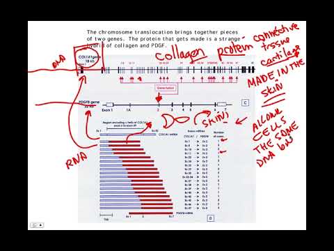
David Champlin explains how a chromosome 17-to-22 translocation associated with dermatofibrosarcoma protuberans (DFSP) joins COL1A1 and PDGFB. The fusion places PDGFB under skin-active COL1A1 regulatory control, causing skin cells to release a growth signal that activates their own PDGF receptors and drives cell-cycle progression.
Key takeaways
- The characteristic chromosome 17;22 translocation joins COL1A1 and PDGFB, a rearrangement that can be identified in biopsy material.
- A gene fusion can alter more than a protein’s coding sequence: the COL1A1 promoter redirects PDGFB expression into skin cells.
- In the cancer mechanism described, skin cells release PDGF and stimulate their own PDGF receptors, forming an autocrine positive-feedback loop.
- PDGF receptor signaling activates phosphorylation pathways that encourage progression through the G1-to-S transition of the cell cycle.
- The example illustrates how cells with the same DNA can produce different proteins because promoters and other regulatory sequences control gene expression.
Chapters
0:00
Microscopically Visible t(17;22) Chromosome Translocation
- Many DNA mutations change a single A, G, C, or T letter and cannot be seen with a microscope; this cancer-associated mutation is a larger chromosome rearrangement.
- Chromosomes 17 and 22 break at specific sites and rejoin, creating a translocation that clinicians can recognize in biopsy cells.
- Humans have 23 chromosome pairs, numbered 1 through 22 plus the sex chromosomes; the characteristic rearrangement involves chromosomes 17 and 22.
2:30
How COL1A1 and PDGFB Form a Cancer-Associated Gene Fusion
- Biochemical analysis identifies breakpoints in PDGFB and COL1A1, a gene that encodes collagen found in connective tissue.
- The rearrangement creates fused genetic material from both genes; Champlin connects this to gene expression and protein production.
- PDGF signaling normally binds a PDGF receptor and can stimulate the cell cycle, while collagen is ordinarily produced by cells such as those in the skin.
5:30
Skin-Specific PDGFB Expression Creates an Autocrine Growth Loop
- Although cells generally share the same DNA, regulatory regions such as promoters control where a gene is expressed; insulin, for example, is produced in the pancreas.
- The COL1A1 promoter normally directs expression in skin; when joined to PDGFB, it drives PDGFB production in skin cells.
- The resulting growth factor is released by exocytosis and binds PDGF receptors on the same cell, creating an autocrine, positive-feedback signal.
- PDGF receptor signaling activates kinase phosphorylation and pushes cells through the G1-to-S cell-cycle checkpoint, promoting growth even without a wound.
Summary, takeaways, and chapters were generated by AI from the video's transcript and may contain errors. The video belongs to its creator, The New Evolution for Everyone.