Protein Structure, BIO105 Introductory Biology, David Champlin, USM
 Watch on YouTube →
Overview
David Champlin explains how the 20 amino acids form proteins whose sequences and interactions determine their three-dimensional structures and functions, using aquaporin as the central example. He connects peptide-bond formation, protein digestion, DNA mutations, and denaturation to explain how a change in amino-acid sequence can alter a protein’s shape and performance.
Key takeaways
- A protein’s primary structure is its specific sequence of amino acids, while interactions among amino acids and with water help produce its higher-order shapes.
- Peptide bonds form through dehydration reactions that release water; digestion uses hydrolysis to break dietary proteins into amino acids.
- Cells build their own proteins from amino acids using DNA instructions, so eating a protein such as plant aquaporin does not supply working aquaporin to human cells.
- A single amino-acid substitution can change protein chemistry and function: sickle-cell hemoglobin substitutes hydrophobic valine for hydrophilic glutamic acid.
- An aquaporin mutation at amino-acid position 192 can replace asparagine with leucine and obstruct the channel, reducing water transport.
- Denaturation disrupts noncovalent interactions and protein shape without breaking the covalent bonds that hold the amino-acid backbone together.
Chapters
0:00
Water Chemistry, Cell Membranes, and Aquaporin’s Shape
- Polar water creates a hydrophilic environment, while amphipathic phospholipids—with hydrophilic and hydrophobic regions—form cell membranes.
- Champlin frames shared cell chemistry and proteins across life as patterns consistent with evolution.
- Aquaporin sits in the cell membrane as a donut-shaped channel built from a folded chain of amino acids.
8:00
Four Levels of Protein Structure and Peptide-Bond Synthesis
- The four structural levels are primary amino-acid sequence, secondary local folds such as spirals, tertiary three-dimensional shape, and quaternary assemblies of multiple protein units.
- Aquaporin’s sequence contains roughly 150 amino acids drawn from the 20 common amino acids; their order helps determine its structure and function.
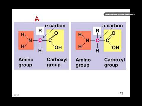
- Amino acids share a backbone but differ in their side chains, which can be hydrophobic, hydrophilic, or amphipathic.
- A dehydration reaction removes water as a peptide bond joins amino acids; hydrolysis reverses the process during protein digestion.
15:00
DNA Mutations Can Change Hemoglobin and Aquaporin Function
- Dietary proteins are digested into amino acids; cells use their own DNA instructions to build human proteins rather than directly reusing, for example, cow insulin or plant aquaporin.
- In sickle-cell anemia, a DNA mutation changes a hemoglobin amino acid from hydrophilic glutamic acid to hydrophobic valine, affecting protein behavior.
- An aquaporin mutation at position 192 changes asparagine to leucine and physically blocks the channel, preventing water from passing normally.
- Inherited aquaporin defects can impair water movement; a mouse example links defective aquaporin to insufficient eye moisture and glaucoma.
21:00
Aquaporin, Osmosis, and Protein Denaturation
- Aquaporin speeds osmosis by allowing water to cross cell membranes rapidly; it does not itself determine whether cells swell or shrink.
- Heat and other harsh conditions can denature proteins by disrupting noncovalent interactions, unfolding their three-dimensional structure while leaving the covalent backbone intact.
- Side-chain interactions help stabilize protein shape, and aquaporin’s donut-shaped units assemble in groups of four as a quaternary structure.
- Aquaporin’s selective water channel illustrates how tertiary structure relates directly to protein function.
Summary, takeaways, and chapters were generated by AI from the video's transcript and may contain errors. The video belongs to its creator, The New Evolution for Everyone.