PDGF and PDGF Receptor, BIO105 Introductory Biology, David Champlin, USM
 Watch on YouTube →
Overview
David Champlin traces platelet-derived growth factor (PDGF) from its production in bone-marrow megakaryocytes to its release at a wound, where it binds the PDGF receptor on nearby skin cells. Receptor signaling promotes passage through the G1-to-S cell-cycle transition and increases cell division; the lecture connects this regulated growth response to mitotic-index assessment of suspicious skin lesions.
Key takeaways
- Bone-marrow megakaryocytes produce PDGF and package it into vesicles that are carried by platelet fragments.
- At a wound, platelets release PDGF alongside clotting factors, coordinating clot formation with a local signal for tissue repair.
- Rapid degradation keeps released PDGF concentrated near the wound, limiting stimulation of cell division elsewhere.
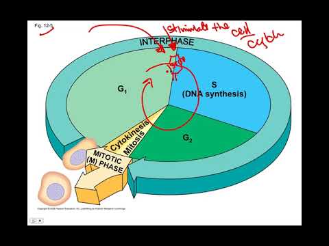
- Binding of PDGF to its specific receptor activates an intracellular kinase and promotes the G1-to-S cell-cycle transition in nearby skin cells.
- Mitotic index is the percentage of cells in mitosis in a tissue sample; a high value in a mole biopsy can indicate rapid growth and warrant clinical concern.
Chapters
0:00
Megakaryocytes Package PDGF into Platelets
- Megakaryocytes in bone marrow produce PDGF and bud off cell fragments that become circulating platelets.
- PDGF is synthesized through the rough endoplasmic reticulum and packaged into vesicles inside the platelet-producing cell.
- Platelets carry both PDGF-containing vesicles and clotting factors; at a wound, their release helps form a clot and delivers PDGF.
3:25
Wound-Localized PDGF Activates Its Receptor
- PDGF released by platelets stays near the wound because it is degraded quickly rather than traveling far through the body.
- Its specific shape enables PDGF to bind the matching cell-surface PDGF receptor, whose intracellular kinase domain relays the signal through protein phosphorylation.
- The signal encourages nearby skin cells to pass the G1-to-S transition, also called the start of the cell cycle, and proceed toward division.
6:30
Mitotic Index Connects Cell-Cycle Activity to Skin Cancer
- Champlin describes mitotic index as the percentage of cells in a tissue sample that are undergoing mitosis; PDGF-driven growth can raise this measure locally.
- A biopsy of a suspicious mole can be examined under a microscope, and a relatively high mitotic index may raise concern about rapidly growing, potentially cancerous tissue.
- The lecture introduces a rare skin cancer, dermatofibrosarcoma protuberans, and leaves its specific cause for a later discussion.
Summary, takeaways, and chapters were generated by AI from the video's transcript and may contain errors. The video belongs to its creator, The New Evolution for Everyone.