Macrophage Movement, BIO105 Introductory Biology, David Champlin, USM
 Watch on YouTube →
Overview
David Champlin explains how the cytoskeleton enables macrophage movement, distinguishing actin microfilaments, tubulin microtubules, and intermediate filaments by their roles. He traces how ATP-powered actin growth and contraction move the cell, while microtubule motor proteins transport recycled integrins to the leading edge so the macrophage can attach to extracellular matrix and advance.
Key takeaways
- Actin microfilaments are built from actin and help change cell shape; microtubules are built from tubulin and provide tracks for intracellular transport.
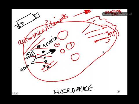
- Macrophage movement combines actin polymerization at the front, which extends the cell membrane, with actin–myosin contraction at the rear, which pushes cell contents forward.
- ATP supplies energy both for actin-based movement and for molecular motors that transport vesicles along microtubules.
- Integrins let a macrophage grip the extracellular matrix, including materials such as collagen, to gain traction as it moves.
- Endocytosis at the rear and exocytosis at the front recycle integrins, maintaining a moving supply of attachment points at the leading edge.
Chapters
0:00
Three Cytoskeleton Components and Their Distinct Roles
- The cytoskeleton is a protein-based framework that supports cell structure and movement.
- Actin microfilaments help cells change shape; actin and myosin also drive muscle contraction.
- Tubulin microtubules act as tracks for moving vesicles, while intermediate filaments help maintain cell and nuclear shape.
4:10
ATP-Powered Actin Remodeling Moves the Macrophage Forward
- Macrophages can crawl out of blood vessels toward bacterial infections and engulf bacteria by phagocytosis.
- Actin microfilaments and myosin contract at the rear, pushing cell contents forward in a motion compared to squeezing toothpaste from a tube.
- ATP hydrolysis to ADP supplies energy for contraction, while ATP-driven actin polymerization at the front pushes the membrane outward.
- Membrane integrins act as attachment points, gripping the extracellular matrix so the cell can push and pull itself forward.
8:20
Microtubules Recycle Integrins from the Rear to the Leading Edge
- After rear integrins release from the extracellular matrix, endocytosis captures them in vesicles.
- ATP-powered molecular motors carry the vesicles along microtubule tracks toward the front of the macrophage.
- Exocytosis returns integrins to the leading edge, where they can attach to the matrix again.
- The coordinated cycle resembles a tank tread: actin changes cell shape, and microtubules reposition adhesion proteins.
Summary, takeaways, and chapters were generated by AI from the video's transcript and may contain errors. The video belongs to its creator, The New Evolution for Everyone.