Hypersensitivity & Autoimmunity 1of2
 Watch on YouTube →
Overview
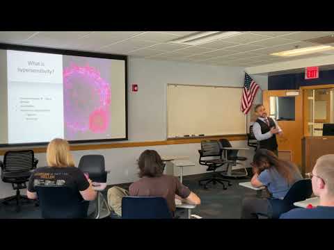
Robert Kellar, Ph.D. explains how adaptive immune responses can become harmful, organizing hypersensitivity into four mechanisms: IgE-mediated allergy, tissue-bound antibody reactions, circulating immune-complex deposition, and T-cell-mediated injury. Examples connect each mechanism to clinical conditions including anaphylaxis, rheumatic fever, myasthenia gravis, Graves disease, glomerulonephritis, multiple sclerosis, type 1 diabetes, and Hashimoto disease.
Key takeaways
- Type I hypersensitivity is an IgE-and-mast-cell response: sensitization during an initial exposure enables rapid mediator release on later exposure, with histamine and leukotrienes contributing to allergy symptoms and airway effects.
- Type II and type III hypersensitivity differ mainly by where antibodies meet antigens: type II antibodies bind antigens on target tissue, whereas type III circulating immune complexes deposit in sites such as kidneys, joints, and vessel walls.
- Group A streptococcal infection can lead to type II rheumatic fever when anti-streptococcal antibodies cross-react with heart or joint tissue, potentially causing carditis and lasting cardiac scarring.
- Autoantibodies can alter receptor function in opposite ways: they block acetylcholine receptors in myasthenia gravis but stimulate thyroid-stimulating hormone receptors in Graves disease.
- Type IV hypersensitivity is driven by T cells rather than antibodies and includes immune injury in conditions such as multiple sclerosis, type 1 diabetes, Hashimoto disease, and contact dermatitis.
- Anaphylaxis can rapidly swell laryngeal tissue and obstruct breathing; severe allergy preparedness centers on emergency treatment and access to epinephrine.
Chapters
0:00
Adaptive Immunity Review: Barriers, Memory, and Vaccines
- First-line defenses include skin, mucous membranes, and other physical or chemical barriers; inflammation is part of innate defense.
- Adaptive immunity uses diverse B and T lymphocytes, central and peripheral tolerance, and memory to respond to a wider variety of pathogens.
- Vaccines create an initial antigen exposure that builds immune memory for a faster, stronger response on later exposure.
4:20
Hypersensitivity as Excessive, Self-Reinforcing Immune Injury
- Hypersensitivity occurs when an immune response to an antigen exceeds what is appropriate and causes tissue damage, as in hives.
- Responses can be triggered or maintained inappropriately and are difficult to control once underway.
- The lecture frames hypersensitivity as a pathological positive-feedback loop, contrasting it with physiological loops such as blood clotting and oxytocin-driven labor.
9:10
Four Hypersensitivity Types: Antibodies, Immune Complexes, and T Cells
- Type I is the IgE-associated allergic response; types II and III involve antibodies, but differ in whether antibodies bind tissue antigens or form circulating complexes.
- Type II examples include incompatible blood transfusions, where ABO and Rh antigens can trigger IgG- and IgM-mediated reactions.
- Type III complexes can lodge in blood vessel walls, joints, or kidneys; type IV is T-cell-mediated and includes transplant rejection.
17:50
Type I Allergy: Sensitization by B Cells and TH2 Cells
- During initial exposure to an allergen such as pollen, a B cell presents antigen and activates a TH2 helper T-cell response.
- B cells produce allergen-specific IgE, which binds to mast cells and sensitizes them for subsequent exposures.
- On repeat exposure, allergen binding activates sensitized mast cells and drives release of inflammatory mediators.
20:26
Mast-Cell Mediators Drive Immediate and Late Allergic Reactions
- Mast-cell degranulation rapidly releases histamine, proteases, and chemotactic factors, causing vasodilation, vascular leakage, and smooth-muscle contraction.
- Bronchial smooth-muscle spasm can make breathing difficult, while histamine-driven blood flow and secretions contribute to watery eyes and nasal symptoms.
- A later phase involves de novo synthesis from arachidonic acid, including prostaglandins and leukotrienes.
24:20
Type I Symptoms: From Hives to Anaphylaxis
- Type I reactions can follow exposure to pollen, dust, pet dander, foods such as peanuts or shellfish, insect venom, or medications such as penicillin.
- Mast-cell mediators and interleukin-13 contribute to tissue inflammation and mucus hypersecretion.
- Anaphylaxis can cause severe laryngeal swelling and airway obstruction; the lecture emphasizes that known severe allergies warrant an emergency response plan and epinephrine access.
29:30
Type II Hypersensitivity: Tissue-Bound Antibodies and Strep Cross-Reactivity
- In type II hypersensitivity, antibodies bind antigens located on cells or tissues; unlike type III, the binding occurs at the target tissue rather than in circulation.
- After group A streptococcal infection, antibodies against streptococcal antigens can cross-react with similar antigens in heart tissue.
- This molecular mimicry can cause inflammation affecting the myocardium, heart valves, and connective tissue.
34:00
Rheumatic Fever and Aschoff Bodies After Streptococcal Infection
- Rheumatic fever can follow streptococcal infection when cross-reactive antibodies inflame the heart and joints; untreated disease may leave lasting cardiac damage.
- Aschoff bodies are associated with rheumatic carditis, and chronic inflammation can scar myocardium and impair heart function.
- The lecture contrasts historical risks before widespread antibiotics with the substantial reduction in severe streptococcal complications after antibiotic treatment became common.
39:57
Type II Autoantibodies: Myasthenia Gravis and Graves Disease
- In myasthenia gravis, antibodies block acetylcholine receptors at the neuromuscular junction, weakening signal transmission and muscle contraction.
- Weakness can affect respiratory muscles, making myasthenia gravis potentially serious rather than merely a loss of strength.
- In Graves disease, stimulatory antibodies activate thyroid-stimulating hormone receptors, increasing T3 and T4 production and causing hyperthyroidism.
45:55
Type III Hypersensitivity: Immune Complexes in Kidneys, Joints, and Vessels
- Type III begins when circulating antibodies bind circulating antigens to form immune complexes that can become trapped in tissues.
- Common deposition sites include kidney filtration structures, synovial joints, and blood vessel walls; complement and neutrophils then drive inflammation.
- The resulting conditions include glomerulonephritis, reactive arthritis, and vasculitis; systemic lupus erythematosus is also introduced as an immune-complex disease.
50:41
Type IV Hypersensitivity: CD4 and CD8 T-Cell Tissue Damage
- Type IV is delayed and cell-mediated, involving CD4 helper T cells, CD8 cytotoxic T cells, and macrophages rather than antibody binding as the defining mechanism.
- CD4 cells recognize antigen presented by cells such as dendritic cells or macrophages and release cytokines that recruit inflammation.
- CD8 T cells can directly kill antigen-bearing target cells; transplant rejection and contact dermatitis from poison ivy or poison oak illustrate this pathway.
53:10
Type IV Disease Examples and the Transition to Autoimmunity
- Multiple sclerosis involves immune injury to central nervous system myelin, while type 1 diabetes involves T-cell destruction of pancreatic beta cells that produce insulin.
- Hashimoto disease destroys thyroid tissue and reduces thyroid hormone production; Graves disease differs because its antibodies stimulate the thyroid receptor.
- Celiac disease is described as T-cell-mediated damage to the intestinal lining, while Crohn disease is presented as a possible type IV condition and IBS is distinguished from it.
- Rheumatoid arthritis is identified as type IV, distinct from antibody-dominated reactive arthritis; the next lecture is set to continue with autoimmune disorders and HIV/AIDS.
Summary, takeaways, and chapters were generated by AI from the video's transcript and may contain errors. The video belongs to its creator, Robert Kellar, Ph.D..